
|
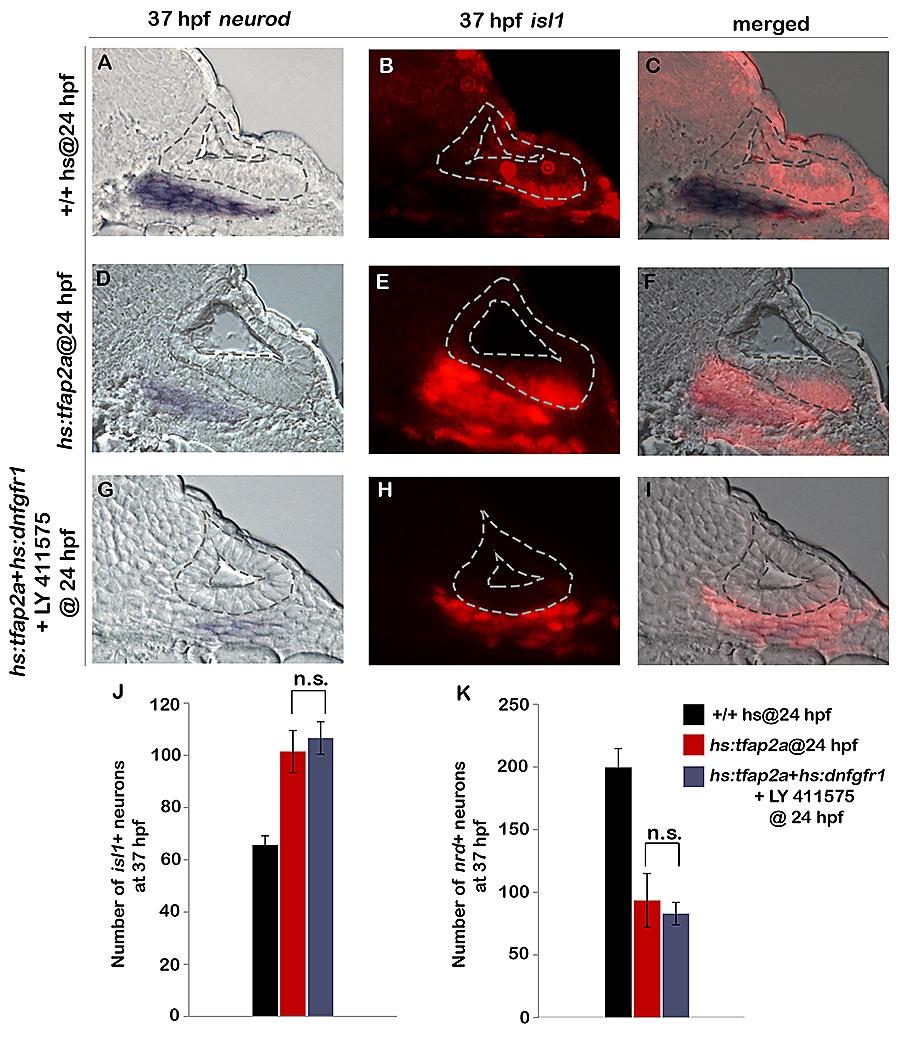
Fig. S4 Inhibition of Notch and Fgf signaling in hs:tfap2a embryos does not enhance the effects of hs:tfap2a activation.
(A-I): Cross-sections at the level of utricular macula (medial to the left, dorsal up) show bright field (A, D, G), fluorescent (B, E, H) and merged (C, F, I) images for neurod (blue) and isl1 (red) in heat-shocked wild-type, hs:tfap2a and LY 411575 treated hs:tfap2a+ hs:dnfgfr1 embryos at 37 hpf. All specimens were treated with 0.3% DMSO and heat-shocked (39°C, 30 minutes) at 24 hpf. (J) Mean and standard deviation of the total number of is1+ neurons at 37 hpf under the conditions indicated in the color key (n = 10–15 specimens each). (K) Mean and standard deviation of the total number of nrd+ neuroblasts at 37 hpf under the conditions indicated in the color key (n = 3–6 ears each, counted from serial sections). Both experimental conditions were significantly different compared to controls. n.s., no statistical difference between the groups indicated in brackets.