
|
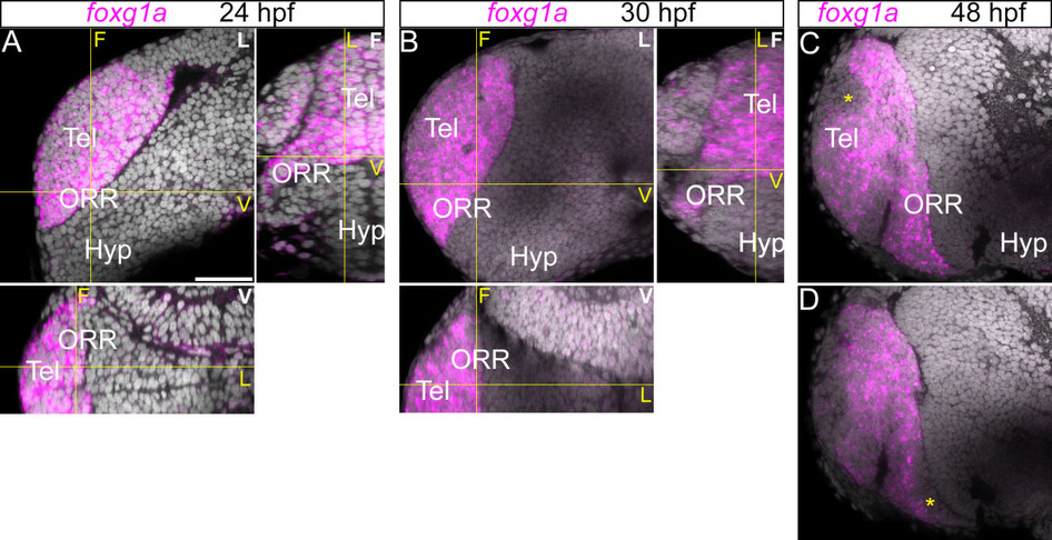
Fig. 8
Expression of foxg1a in the telencephalic region.
Anterior forebrain region following foxg1a in situ hybridization and DAPI staining (gray) at 24 (A), 30 (B) and 48hpf (C). (A–B): A single confocal plane of a lateral view (left top panel; indicated “L” in white) and the frontal (right top; indicated “F” in white) and ventral (bottom panel; indicated “V” in white) views reconstructed using Z-projections of the lateral images. Corresponding section levels are indicated in yellow lines and yellow letters. The foxg1a is expressed in the entire region antero-dorsal to the optic recess. (C–D): A single confocal plane of two different lateral views at 48hpf (C more medial and D more lateral). The expression of foxg1a is reduced at 48hpf in some territories (*). Scale bar = 50µm.