
|
Fig. 3
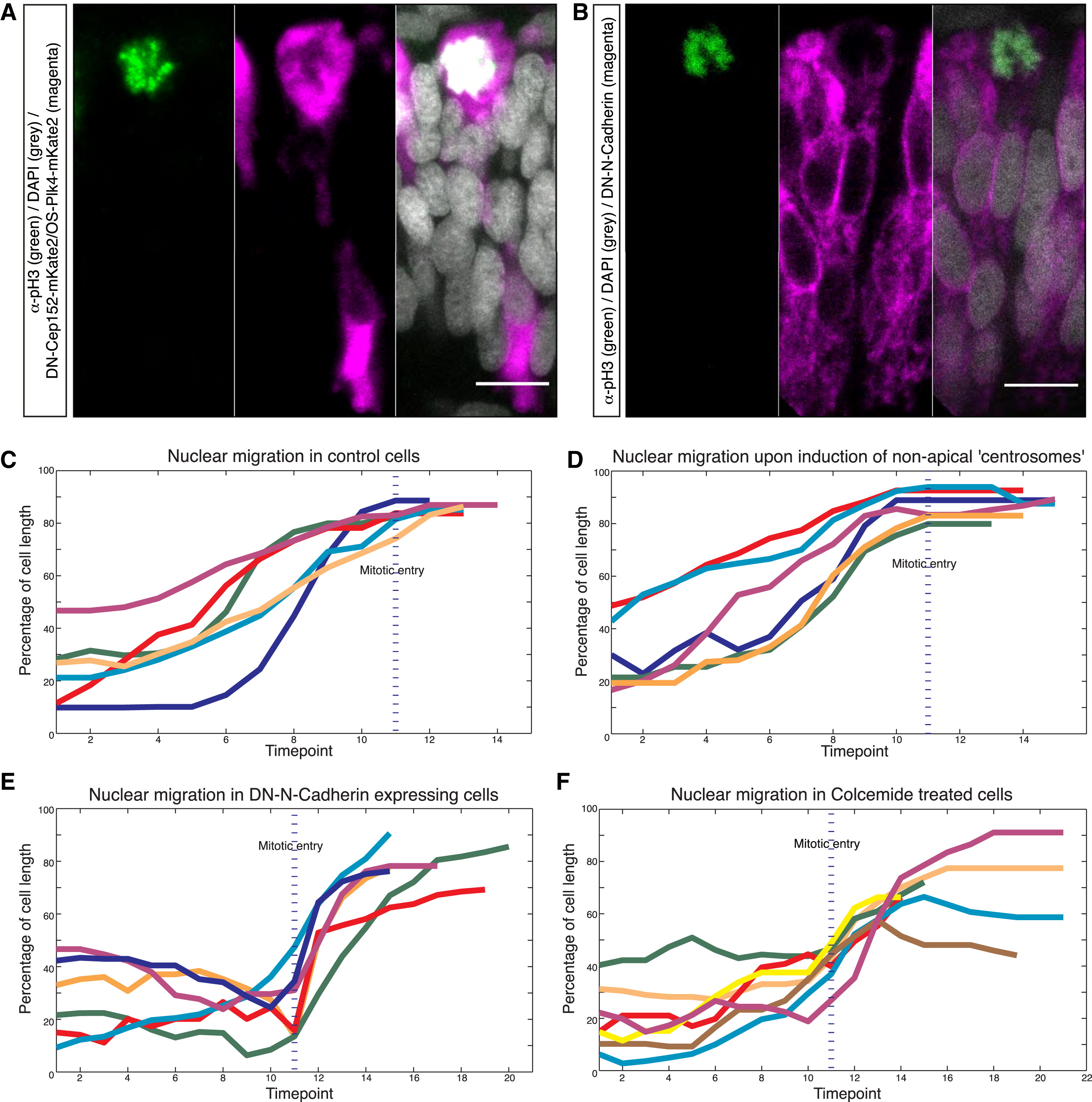
Apical IKNM Persists Despite Nonapical Mitotic Entry
(A and B) pH3 stainings (green) of embryos expressing HS-DN-Cep152-mKate2/HS-OS-Plk4-mKate2 (A) or DN-N-Cadherin (B) (both magenta).
(C–F) Tracks of nuclear position around mitosis in (C) control cells, (D) cells featuring nonapical centrosomes, (E) cells expressing DN-N-Cadherin, (F) cells treated with 100µM colcemide. Time shown in time points (tp). 1 tp = 5 min. Nuclear position was tracked 10 tp (= 50 min) prior to and 10 tp after mitotic entry or until division. Nuclear position has been measured from the base of the nucleus to the basal lamina and was normalized with respect to cell length. Each track represents a single nucleus; tp of mitotic entry is marked with blue line (always at tp = 11).
Scale bars represent 10 µm.
Reprinted from Developmental Cell, 32(2), Strzyz, P.J., Lee, H.O., Sidhaye, J., Weber, I.P., Leung, L.C., Norden, C., Interkinetic Nuclear Migration Is Centrosome Independent and Ensures Apical Cell Division to Maintain Tissue Integrity, 203-19, Copyright (2015) with permission from Elsevier. Full text @ Dev. Cell