
|
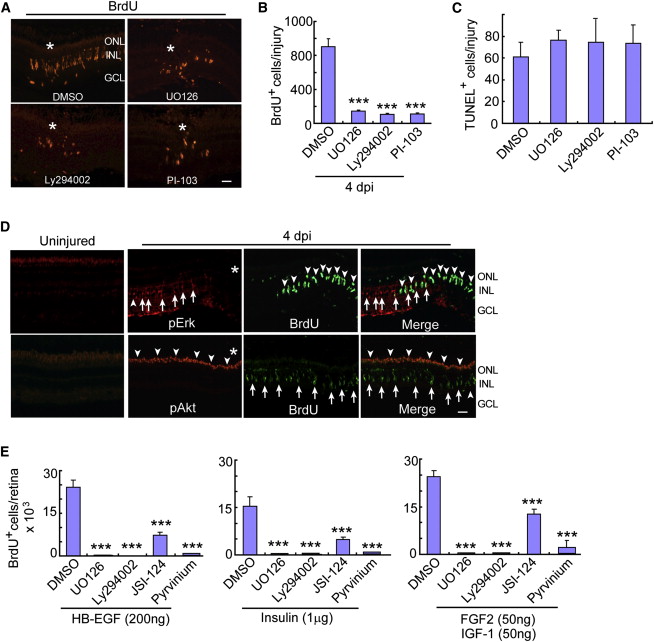
Fig. 3
Mapk/Erk, PI3K/Akt, β-Catenin, and Jak/Stat Signaling Regulate Injury and Growth Factor-Stimulated MG Proliferation
(A and B) Mapk/Erk inhibitor (UO126) or PI3K inhibitors (Ly294002 or PI-103) reduce the number of BrdU+ progenitors at 4 dpi. Asterisks identify the injury site. Scale bar represents 50 µm. Error bars represent SD; p < 0.001; n = 6.
(C) TUNEL assay shows Mapk and PI3K inhibitors have no effect on cell death.
(D) pErk and pAkt immunofluorescence in uninjured and injured (4 dpi) retinas. Asterisks identify the injury site. In the pErk panels, arrows point to pErk+ MG and arrowheads point to BrdU+ cells. In pAkt panels, arrowheads point to pAkt+ cells, while arrows point to BrdU+ cells. Scale bar represents 50 µm.
(E) In the growth factor stimulated, but uninjured retina, BrdU+ progenitor formation is suppressed by inhibiting Mapk (UO126), PI3K (Ly294002), Jak/Stat (JSI-124) or β-catenin (pyrvinium) signaling. Error bars represent SD; p < 0.001; n = 5.
See also Figures S2–S4.