
IMAGE
Fig. 3
- ID
- ZDB-IMAGE-150330-25
- Genes
- Antibodies
- Publication
- Miller et al., 2015 - Neurobeachin Is Required Postsynaptically for Electrical and Chemical Synapse Formation
- All Figures
- Figures for Miller et al., 2015
Image
|
Figure Caption
Fig. 3
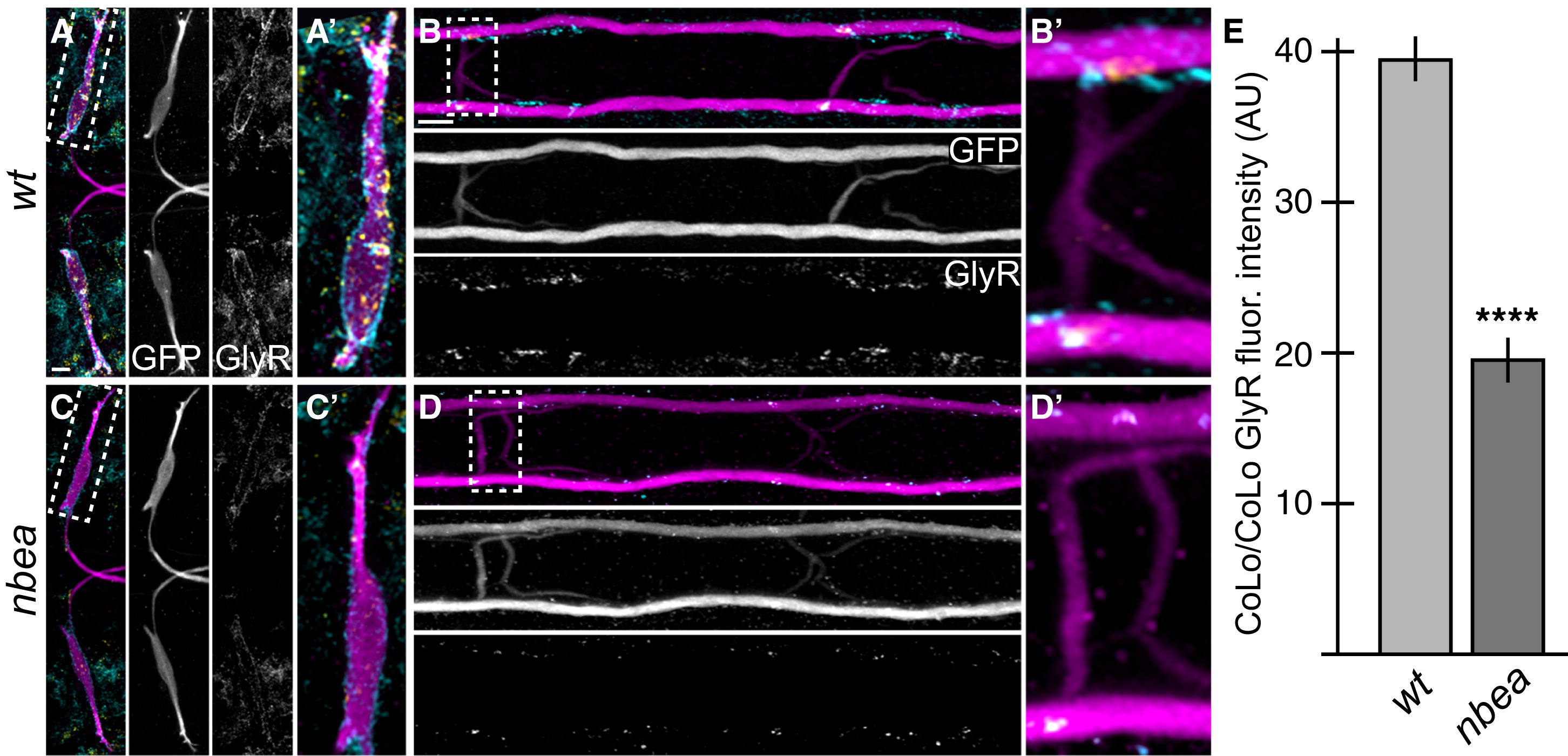
Glycinergic Synapses Are Disrupted in nbea Mutants
Larvae were stained for GFP (magenta), Cx36 (yellow), and glycine receptor (GlyR, cyan). Individual GFP and GlyR channels are shown in neighboring panels.
(A–D) The GlyR staining found on M dendrites (A) and CoLo/CoLo synapses (B) is diminished in nbea mutant animals (C and D).
(E) Quantitation of the amount of GlyR in wild-type and mutant CoLo/CoLo synapses. See Figure 1A for circuit diagram. Graphs represent data as mean ± SEM. p < 0.0001 compared to control.
Associated experimental statistics are found in Table S2.
Figure Data
Acknowledgments
This image is the copyrighted work of the attributed author or publisher, and
ZFIN has permission only to display this image to its users.
Additional permissions should be obtained from the applicable author or publisher of the image.
Full text @ Curr. Biol.