
|
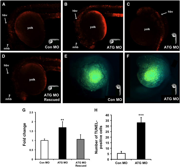
Fig. 6
Abrogation of glut2 enhances cell death mainly in the head region. To assay for cell death, embryos injected with control morpholino (Con MO; A), ATG morpholino (ATG MO; B and C), and ATG MO+rat GLUT2 mRNA (ATG MO Rescued; D) were stained with the vital dye acridine orange. To assay for apoptosis, embryos injected with Con MO (E) and ATG MO (F) were analyzed by TUNEL assay. At 24 hpf, there was an overall increase in cell death and apoptosis primarily localized in the hindbrain region (B, C, and F). Fluorescent signal analysis (G) and counting of TUNEL-positive cells (H) confirmed a significant increase in cell death and apoptosis, respectively, in ATG morphants which appears reverted in rescued embryos (G). * indicates significant differences compared with the Con MO injected embryos (*P<0.01; ***P<0.001). Hindbrain ventricle (hbv), midbrain/hindbrain boundary (mbh), telencephalon (t). In the images (A–F), the position of the embryos is indicated by a representation of a zebrafish embryo.