
|
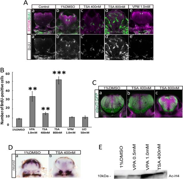
Fig. 2
The HDAC inhibitor TSA mimics the effect of VPA on cell proliferation in embryonic zebrafish. A: TSA-treated brains were stained with anti-BrdU antibody. Number of BrdU-positive cells increased in a dose-dependent manner, and the area of BrdU-positive cells expanded in the TSA-treated brain. B: Number of BrdU-positive cells per section. C: Tg(elavl3:GFP) fish were treated with TSA. GFP-positive area decreased in the TSA-treated embryo, similar to the VPA-treated embryo. D: In situ hybridization of 400 nM TSA-treated embryos with neurod probe. Expression of neurod decreased in the TSA-treated brain. E: Acetylated histone H4 (Ac-H4) level was analyzed by Western blot analysis in brain homogenates from zebrafish embryos treated with indicated drugs. Increased Ac-H4 level was observed in 1.0 mM VPA and 400 nM TSA-treated samples. **P < 0.01, ***P < 0.001