
Image
|
Figure Caption
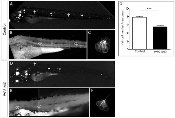
Fig. 7
Decreased hair cell number of PLL neuromast in PrP2-MO.
A–C. In control embryos at 48 hpf, Brn3-GFP fluorescence labels hair cells of neuromasts and ear. High magnification of the first 3 neuromasts and zoom of the first neuromast show 8 hair cells. D–F. Morphants displays reduced number of neuromasts. High magnification shows smaller hair cells number. G. Quantification indicates significant reduction of hair cells/neuromast, independently of the neuromast position (mean hair cell number: 5.5±0.4, n = 36, compare to control 7.9±0.2, n = 20, p<0.001).
Figure Data
Acknowledgments
This image is the copyrighted work of the attributed author or publisher, and
ZFIN has permission only to display this image to its users.
Additional permissions should be obtained from the applicable author or publisher of the image.
Full text @ PLoS One