
|
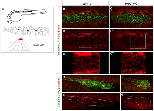
Fig. 6
Delocalisation of E-cadherin and beta-catenin in primordium cells in absence of PrP2.
A. Schematic representation of claudinB-GFP embryo at 30 hpf, primordium structure, and transversal view of a rosette showing the level of confocal focus plan. B–B′′. In control embryos at 30 hpf, E-cadherin is expressed at the membrane level at the basal side of the primordium. C–C′′. In morphant embryos, E-cadherin is observed in the cytoplasm and cellular membrane at basal and apical levels. The staining pattern is more punctuated in morphant compared to control. D, E. In control embryos, beta catenin expression is observed at the membrane level and at the center of rosette structures. F, G. In morphant, small rounded primordium shows a membrane homogeneous pattern with no rosette.