
Image
|
Figure Caption
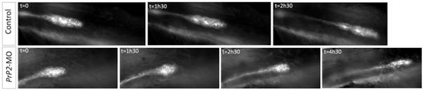
Fig. 5
Loss of cell-cell contact and disorganization of collective migration of the primordium.
Select time points from time-lapse recording of primordium migration in claudinB-GFP embryos. In control embryos, primordium migration is continuous, with a neuromast deposition (time point 2h30) and out of view at the time point 4h30 (not shown). In contrast in one PrP2 morphant example, representative of the observed phenotypes, primordium migration shows a progressive rounded shape and arrest. See also Movies S6 and S7.
Figure Data
Acknowledgments
This image is the copyrighted work of the attributed author or publisher, and
ZFIN has permission only to display this image to its users.
Additional permissions should be obtained from the applicable author or publisher of the image.
Full text @ PLoS One