
|
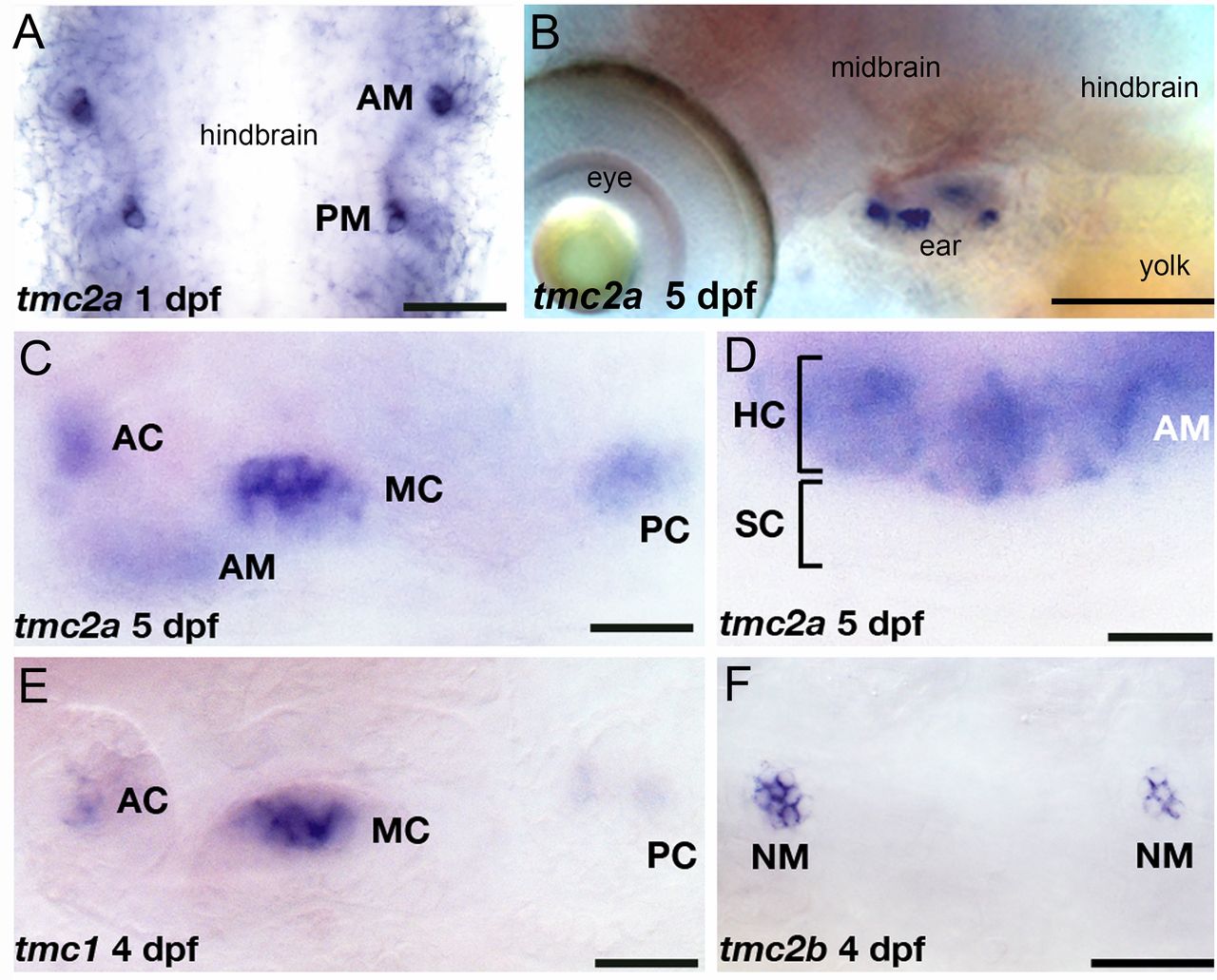
Fig. 3
Embryonic and larval expression pattern of the tmc1/2 genes. In situ hybridization with specific probes for each transcript was performed on stages 1–5 dpf. Expression of tmc2a at 1 dpf (A, flat mount) and 5 dpf (B). Higher magnification view of tmc2a expression in the inner ear (C) and anterior macula at 5 dpf (D). Label is restricted to the upper hair cell layer of the neuroepithelium. (E) Expression of tmc1 at 4 dpf (focal plane: cristae). (F) Expression of tmc2b in neuromasts, 4 dpf. AC, anterior crista; AM, anterior macula; HC, hair cells; MC, medial crista; NM, neuromast; PC, posterior crista; SC, supporting cells. [Scale bars: 50 μm (A); 130 μm (B); 22 μm (C and E); 12 μm (D); 43 μm (F).]