
|
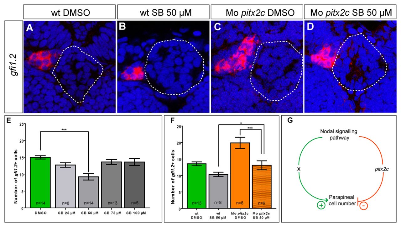
Fig. 4
Suboptimal Nodal signalling reduces parapineal cell number. (A-D) Single confocal sections of the pineal complex showing in situ hybridisation against gfi1.2 in mock-treated, 50 μM SB431542-treated, mock-treated/pitx2c morphant and 50 μM SB431542-treated/pitx2c morphant embryos at 72 hpf. The outline of the pineal gland is highlighted with a dotted line. (E) Counts of cells expressing gfi1.2 in mock-treated and SB431542-treated embryos. The number of gfi1.2-positive cells is significantly decreased only when embryos are treated with 50 μM SB431542; ***P<0.0.001 using a t-test. Error bars represent s.e.m. (F) Counts of cells expressing gfi1.2 in mock-treated and SB431542-treated embryos injected with pitx2c morpholino. Whereas DMSO-treated morphant embryos leads to an increase in parapineal cell number, morphants embryos treated with 50 μM of SB431542 display wild-type numbers of parapineal cells; *P<0.05, ***P<0.0.001 using a t-test. Error bars represent s.e.m. (G) Model for the antagonism between Nodal and Pitx2c. Whereas Nodal activates pitx2c expression that subsequently limits parapineal cell numbers, in parallel a second target of Nodal signalling (X) promotes an increase in parapineal cell numbers. The balance between these two Nodal effectors provides fine control over the maximum cell number of the parapineal.