
|
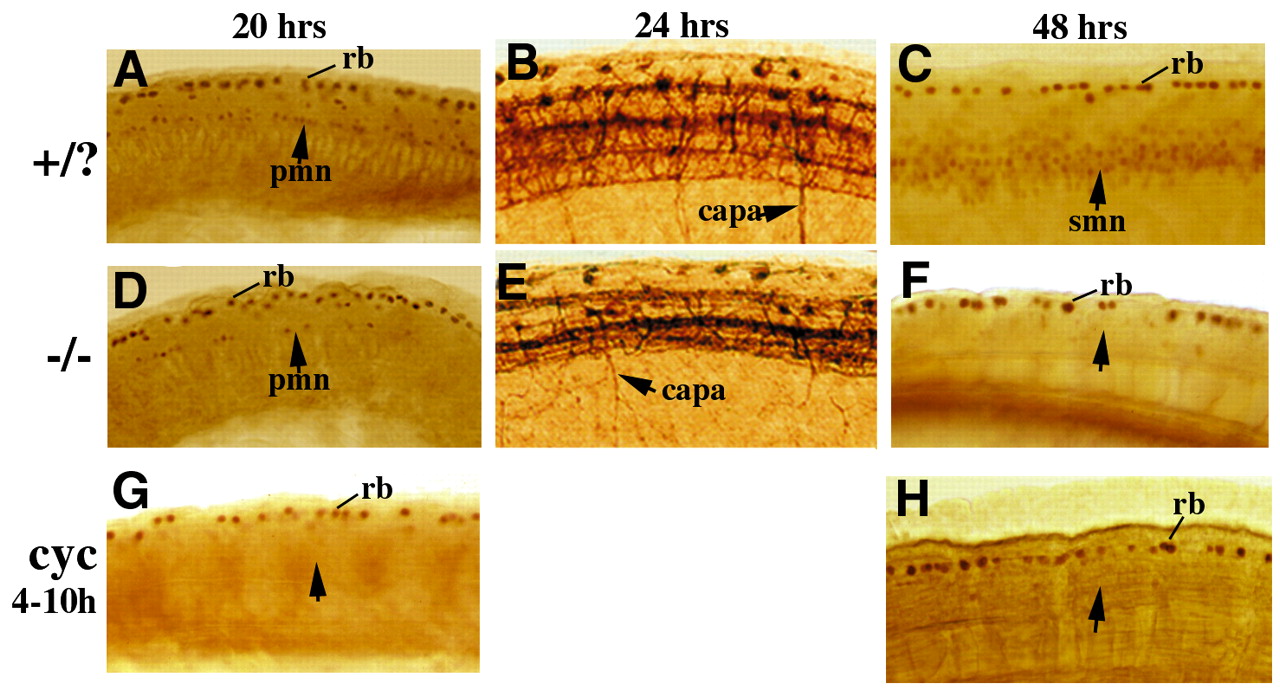
Fig. 5 Maternal and zygotic smo function in MN development. (A,D,G) Lateral views of islet-1/2 antibody staining showing MNs in wild-type (A), smo mutant (D) and cyclopamine treated wildtype (G) embryos at 20 hpf. Mutant embryos have a decreased number of PMNs (arrow). The decrease becomes more severe in the caudal spinal cord. Cyclopamine treatment during gastrulation completely eliminates PMN formation (G). (B,E) Flattened lateral views of acetylated α-tubulin staining showing axons of primary neurons at 24 hpf. The axons of caudal PMN (CaP) in each hemisegment extend down (B, arrow), while CaP axons in the mutant branch and extend randomly (E, arrow). (C,F,H) Lateral views of Islet- 1/2 antibody stained embryos at 48 hpf. A large number of SMNs are seen in the wild-type embryos (C, arrow), whereas few can been seen in the mutant (F, arrow). The PMNs disappear almost completely. Treatment of embryos with cyclopamine during gastrulation in wild-type embryos also leads to the absence of SMN formation (H, arrow). capa, caudal primary motoneuron axons; pmn, primary motoneuron; smn, secondary motoneuron; rb, Rohon-Beard neurons.