
|
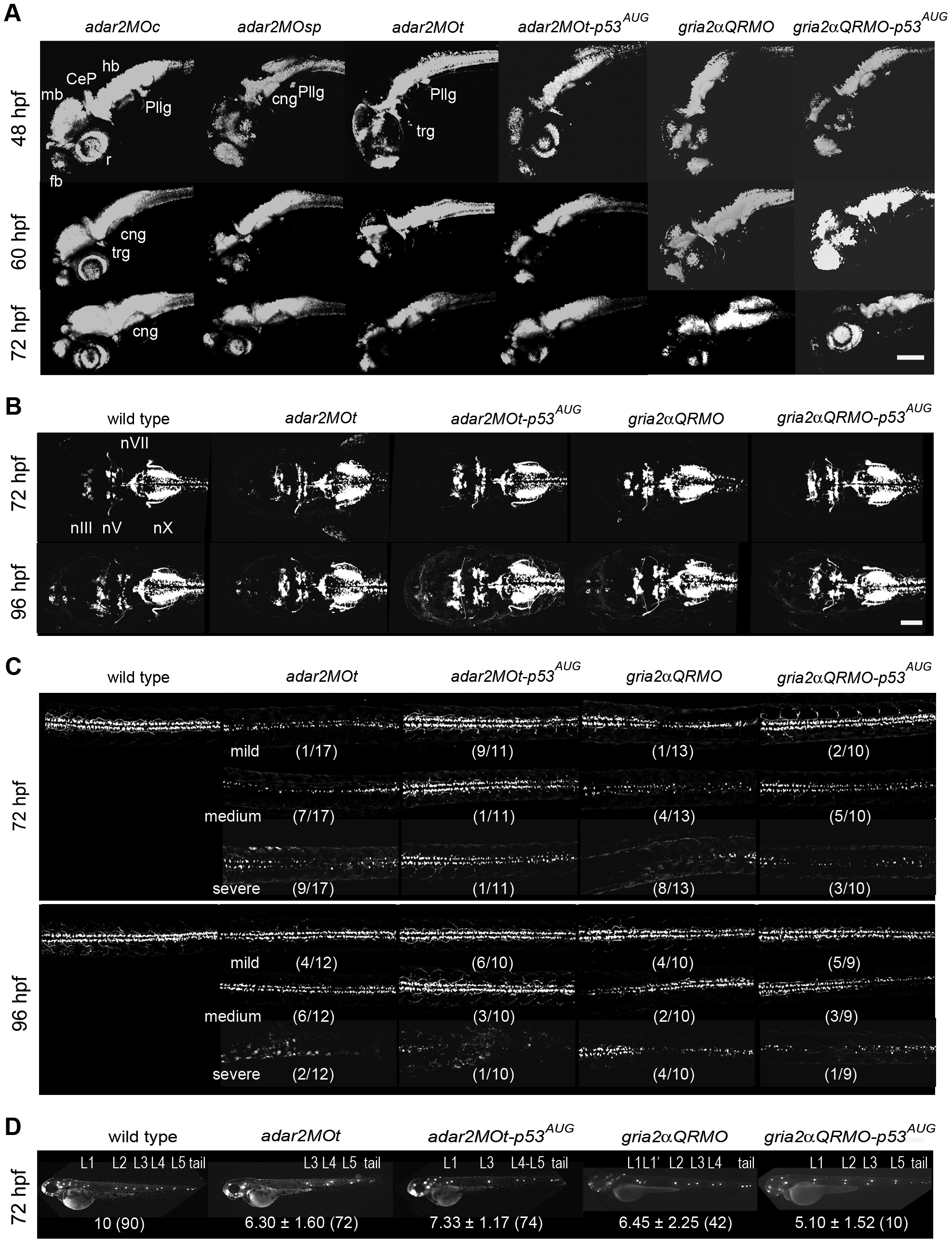
Fig. 6
Defective development of the nervous system in the hypo-Q/R-editing morphants.
(A) The development of early and mature neurons. Confocal microscopic observations of the kaede fluorescence in Tg(HuC:kaede) receiving morpholino injections. The kaede-expressing domain is reduced in the brain of hypo-Q/R-editing morphants. CeP, cerebellar plate; cng, cranial ganglion; fb, forebrain; hb, hindbrain; pllg, posterior lateral line ganglion; r, retina; trg, trigeminal neuron. (B) The development of cranial motor neurons. Confocal microscopic observations of the GFP in the heads of Tg(isl1:GFP) receiving morpholino injections. The cranial motor neurons are only mildly affected. nIII; oculomotor nuclei; nV; trigeminal nuclei; nX, vagus nuclei. (C) The development of spinal motor neurons. Confocal microscopic observations of the GFP in the trunks of Tg(isl1:GFP) receiving morpholino injections. The spinal motor neurons dorsal to the yolk extension are shown. The effects of morpholino treatments were classified into three groups by relative density of motor neuron in each treatment. The numbers in parenthesis indicate the numbers of larvae in a class over all the observed larvae. Scale bar represents 100 μm. (D) The development of lateral line neuromasts. Epifluorescent microscopic observations of the lateral line neuromasts stained by Di-4-Asp [48]. L1-L5 are the posterior lateral line neuromasts, and tail represents the tail neuromasts. L12 is the secondary PLL neuromast. The average ± s.d. (number of larva) of the primary lateral line neuromasts are shown below. Larvae without tail neuromast were excluded from the analysis.