
|
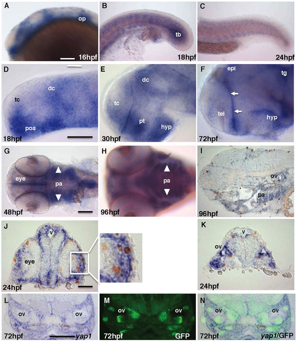
Fig. 1
(A–C) yap1 is expressed in the brain, somites and tail bud until mid-somitogenesis and its expression in the posterior body declines later on. (D–F) yap1 expression become more restricted to the ventricular zones (proliferative region, arrows) in the brain as the embryo matures. (G–I) Staining of yap1 in pharyngeal arch (arrowhead) at 48–96 hpf. (J), (K) Most proliferative cells detected by anti-pH3 antibody (brown, cell nucleus) are yap1-positive (blue) in cross-section of 24 hpf and (I) sagittal-section of 96 hpf embryos. L-N – cross-sections at the mid-hindbrain (ear) level of 72 hpf SqET33-mi60A transgenic larvae (lnfg, progenitors of sensory cells). (L) yap1 in situ hybridization; (M) GFP expression; (N) composite yap1 in situ hybridization/GFP expression. (A–F) – lateral view, (G), (H) – ventral view. Scale bar = 40 μm. Abbreviations: dc, diencephalon; epi, epiphysis; hyp, hypothalamus; op, otic placode; pa, pharyngeal arches; poa, preoptic area; pt, prethalamus; tb, tail bud; tc, telencephalon; tg, tegmentum.