IMAGE

Fig. 5

ID
ZDB-IMAGE-140808-5
Genes
Antibodies
Source
Figures for Monteiro et al., 2014
Image
Figure Caption

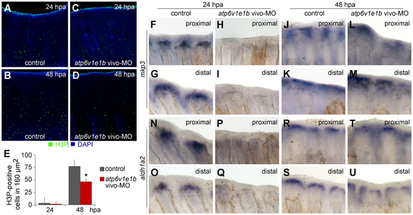
Fig. 5 V-ATPase is required for blastema cell proliferation and expression of mkp3 and aldh1a2.

(A–D) Immunohistochemical detection of proliferating cells (H3P-positive) 24 h (A, C) and 48 h (B, D) after proximal amputation. (A, B) control fin; (C, D) V-ATPase knocked down fin. (E) Quantification of H3P-positive cells in the blastema. *statistical significant results (p<0.05). (F–U) In situ hybridization for mkp3 (F-M) and aldh1a2 (N-U), after proximal amputation (F, H, J, L; N, P, R, T) and distal amputation (G, I, K, M; O, Q, S, U): (F–G; N–O) control fin, 24 hpa; (H–I; P–Q) V-ATPase knocked down fin, 24 hpa; (J–K; R–S) control fin, 48 hpa; (L–M; T–U) V-ATPase knocked down fin, 48 hpa. Control fin: caudal fin from AB wild type fish, non-treated. V-ATPase knockdown procedure: atp6v1e1b vivo-MO delivered to the caudal fin of atp6v1e1bhi577aTg/+ (AB) fish, 2 hpa. hpa: hours post amputation. For each panel, n = 3.

Figure Data
Acknowledgments
This image is the copyrighted work of the attributed author or publisher, and ZFIN has permission only to display this image to its users. Additional permissions should be obtained from the applicable author or publisher of the image. Full text @ PLoS One