
|
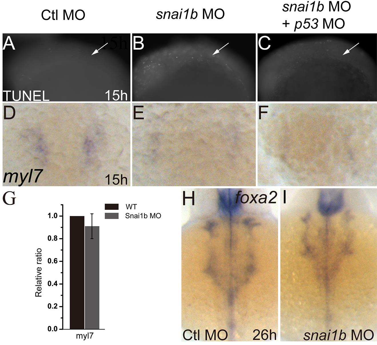
Fig. S3 Heart defects induced by snai1b knockdown are not due to apoptosis or endodermal defects. (A–C): TUNEL assay shows increased apoptosis in the snai1b MO injected embryos (B) compared to controls (A). Co-injection of p53 MO inhibits apoptosis (C) at 15 hpf. White arrows: cardiac precursor regions. (D–F): myl7 expression was decreased in snai1b MO injected embryos (E) compared to controls (D), and p53 MO did not rescue myl7 expression at 15 hpf. (G): q-PCR showed the myl7 expression do not change at 15 hpf (H, I): endoderm formation was not influenced, as judged by the expression of the endoderm marker foxa2 at 26 hpf.