
Image
|
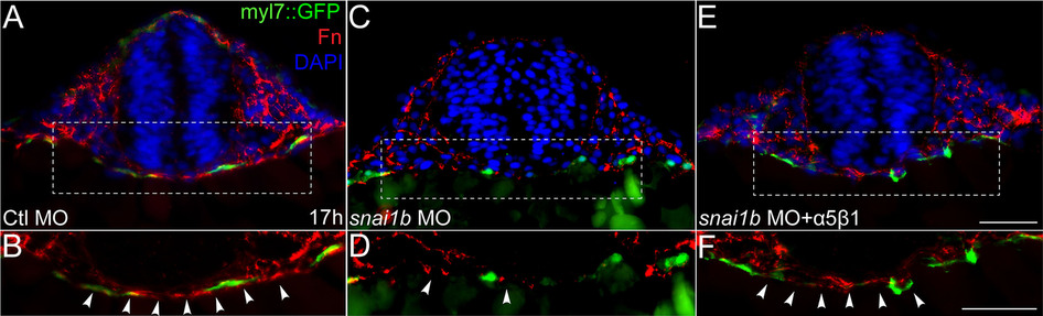
Figure Caption
Fig. 3
Transverse sections of myl7::GFP (green) transgenic embryos immunostained for Fn (red). DAPI (blue) staining indicates nuclei. (A, B): Fn fibrils around myocardial precursors and across the midline in control embryos. (C, D): Fn levels decrease in snai1b MO injected embryos. (E, F): Fn levels are restored in embryos injected with α5β1 integrin protein. (B, D, and F): Magnified view of dashed rectangle of A, C, and E, respectively. Scale bars: 50µm.
Figure Data
Acknowledgments
This image is the copyrighted work of the attributed author or publisher, and
ZFIN has permission only to display this image to its users.
Additional permissions should be obtained from the applicable author or publisher of the image.
Full text @ Sci. Rep.