
Image
|
Figure Caption
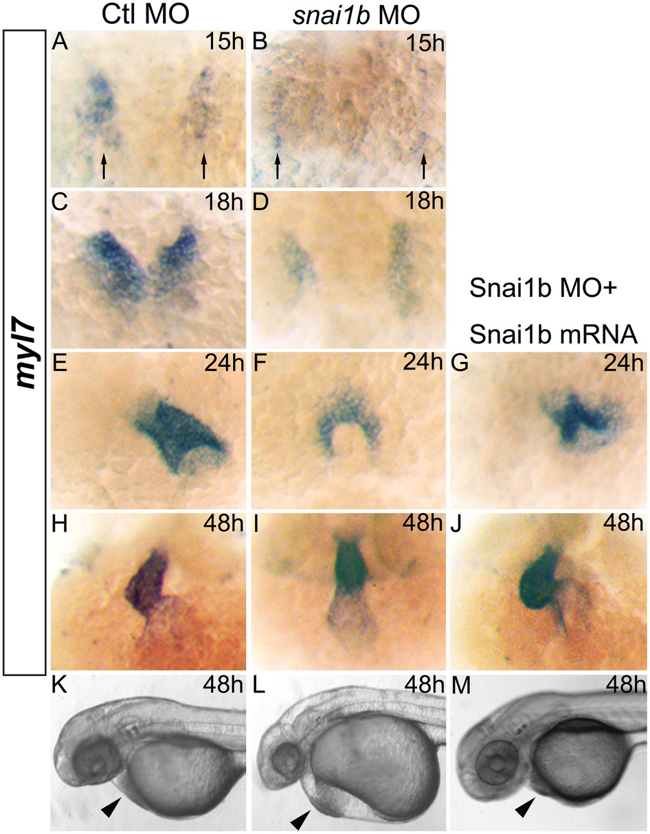
Fig. 2 (A–J): myl7 expression marking myocardial precursor cells in zebrafish embryos. (A, C, E, H): embryos injected with Ctl MO develop normally at 15, 18, 24, and 48hpf. (B, D, F, I): embryos injected with snai1b MO display cardiac fusion delays (B, D, F) and band-like heart shape at 48hpf (I). Myl7 expression (black arrows) is reduced at 15 and 18hpf (B, D). (G, J): embryos injected with both snai1b MO and mRNA display partial rescue of heart defects. (K–M): rescue of heart defects (arrowhead) in embryo injected with snai1b MO and mRNA at 48hpf.
Figure Data
Acknowledgments
This image is the copyrighted work of the attributed author or publisher, and
ZFIN has permission only to display this image to its users.
Additional permissions should be obtained from the applicable author or publisher of the image.
Full text @ Sci. Rep.