
|
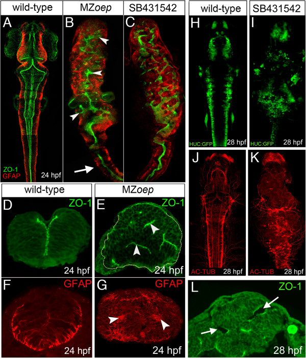
Fig. 1 MZoep mutants have aberrant neural tube organization. (A) Projection of confocal z-series showing dorsal view of brain and anterior spinal cord. The midline ventricle is lined by ZO-1 expression (green) and the basal regions of the neuroepithelium are lined by the GFAP expression (red). (B) Projection of confocal z-series showing dorsal view of brain and anterior spinal cord from MZoep mutant. Both apical ZO-1 and basal GFAP expression show extensive disruption to neural tube morphology in brain regions (arrowheads) but appear relatively normal in anterior spinal cord (arrow). (C) Projection of confocal z-series showing dorsal view of brain and anterior spinal cord from embryo treated with the Nodal inhibitor SB-431542. (D,E) Transverse sections show the normal single midline domain of ZO-1 appears discontinuous and more randomly oriented in MZoep embryo. (F,G) The basal marker GFAP is expressed in ectopic foci deep from the surface of the MZoep neural primordium. (H,I,J,K) Neurons labeled with tg(HUC-GFP) (green) and their axons labeled with Ac-tub (red) are present but disorganized in the Nodal-defective embryo brains. (L) By 28 hpf ectopic ventricles (arrowed) have opened up in the MZoep brains. Ac-tub, anti-acetylated tubulin antibody; GFAP, glial fibrillary acidic protein; hpf, hours post fertilization; MZoep, maternal-zygotic one-eyed pinhead; ZO-1, zonula occludens 1.