
|
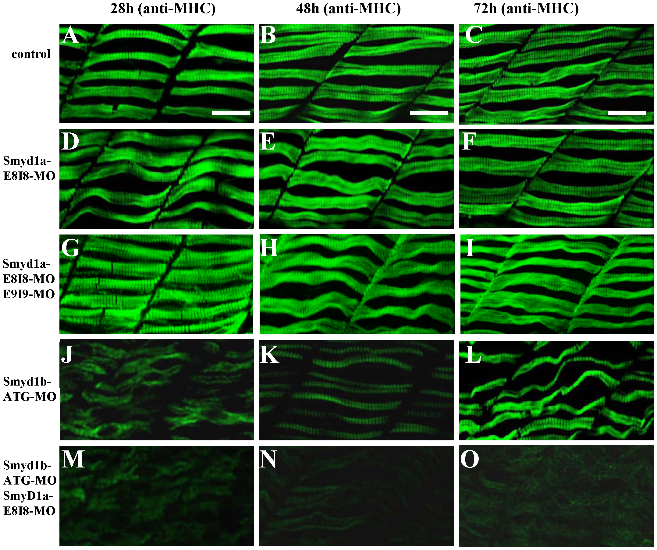
Fig. 3
The effect of smyd1a and/or smyd1b knockdown on myosin thick filament organization in skeletal muscles.
Zebrafish embryos injected with smyd1b MO or smyd1a MO or both were fixed at 28, 48 and 72 hpf. Myosin thick filament organization was analyzed by immunostaining with the F59 antibody which recognizes the myosin heavy chain (MHC) in slow muscles, and followed by FTIC-labeled secondary antibody. The images represent side view of trunk muscles around segment 10. A–C. Lateral view of thick filament organization in slow muscle fibers of control-MO injected embryos at 28 (A), 48 (B) and 72 (C) hpf. D–F. Lateral view of thick filament organization in slow muscle fibers of smyd1a E8I8-MO injected embryos at 28 (D), 48 (E) and 72 (F) hpf. G–I. Lateral view of thick filament organization in slow muscle fibers of smyd1a E8I8-MO and E9I9-MO co-injected embryos at 28 (G), 48 (H) and 72 (I) hpf. J–L. Lateral view of thick filament organization in slow muscle fibers of smyd1b ATG-MO injected embryos at 28 (J), 48 (K) and 72 (L) hpf. M–O. Lateral view of thick filament organization in slow muscle fibers of smyd1a E8I8-MO and smyd1b ATG-MO co-injected embryos at 28 (M), 48 (N) and 72 (O) hpf. Scale bars: 20 μm in A–C.