
|
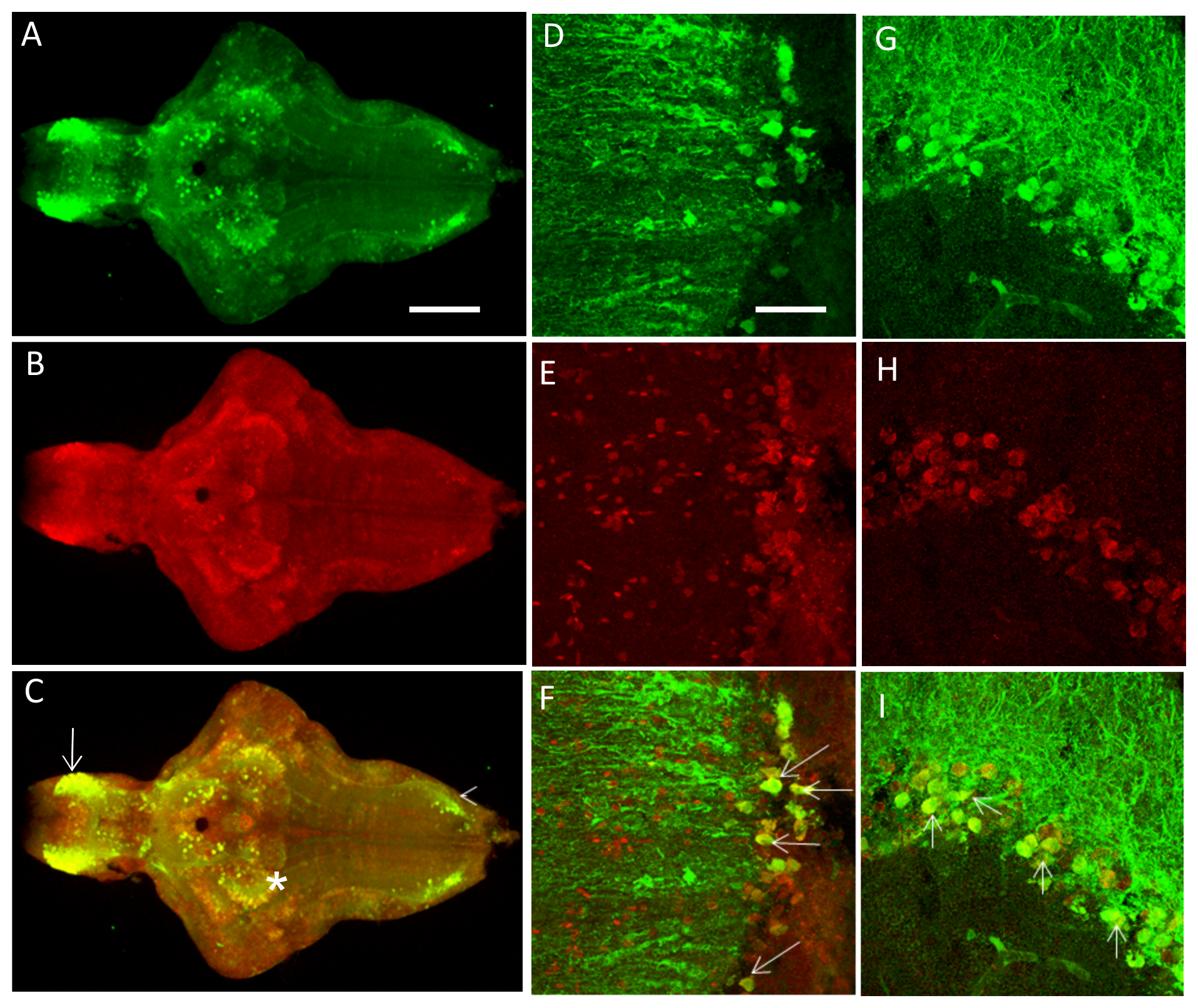
Fig. 2
A-C. The PINK1-ir colocalizes with the GFP expression pattern in the brain of 7-dpf fish from the transgenic line in most parts of the brain. Colocalization was visualized in the telencephalon (arrow), the thalamic region (*), and the rhombencephalon (arrowhead).
D-L. Representative higher magnification images from adult brain sections (25 μm) for PINK1-ir and GFP-ir. D-F represents the telencephalic region in the saggital plane. G-I represents the thalamic region, and J-L represents the rhombencephalic region from the coronal plane. GFP – Green fluorescent protein, PINK1 – PTEN-induced putative kinase 1, ir – Immunoreactivity.