
Fig. 2
- ID
- ZDB-IMAGE-140730-118
- Publication
- Weber et al., 2013 - Characterization of light lesion paradigms and optical coherence tomography as tools to study adult retina regeneration in zebrafish
- All Figures
- Figures for Weber et al., 2013
|
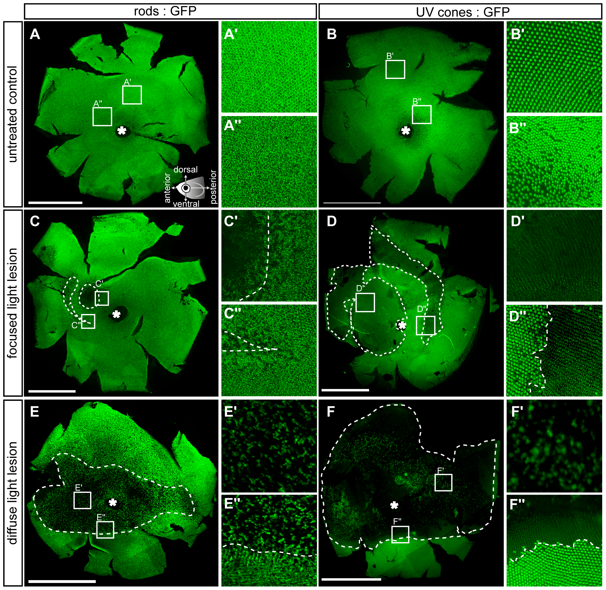
Fig. 2
Flatmounted retina samples of lesioned GFP transgenic reporter fish.
Rods (left column) are labelled with the rh1:GFP and UV cones (right column) with the opn1sw1:GFP reporter fish, respectively. A, B: Vitreal view of untreated control eyes. Insets show magnified images indicated in the overviews. Illustration showing the orientation of flatmounted samples as vitreal view with dorsal (superior) orientation to the top. C-C3: 3 days post focused light lesions in rh1:GFP. D-D3: 3 days post focused light lesions in opn1sw1:GFP. E-E3: 3 days post diffuse light lesions in rh1:GFP. F-F3: 3 days post diffuse light lesions in opn1sw1:GFP. The asterisk in A–F marks the optic nerve head. Scale bars represent 1 mm.