
Fig. 12
- ID
- ZDB-IMAGE-140730-117
- Publication
- Weber et al., 2013 - Characterization of light lesion paradigms and optical coherence tomography as tools to study adult retina regeneration in zebrafish
- All Figures
- Figures for Weber et al., 2013
|
Fig. 12
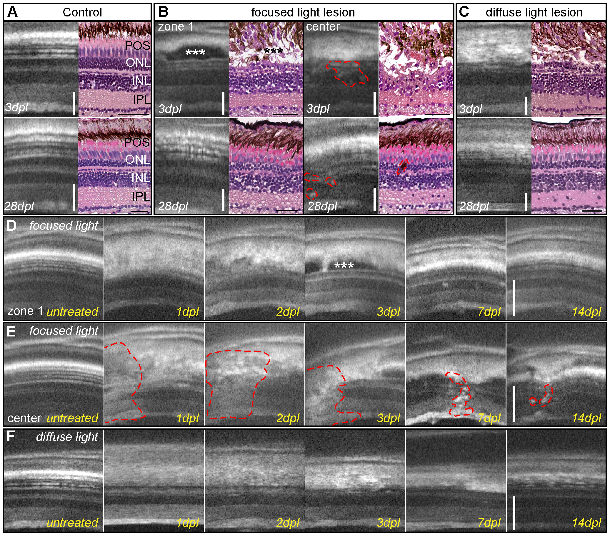
Imaging of light lesions with SD-OCT.
A: Comparison of OCT images to HE staining, showing nuclear layers in dark shades and plexiform layers in grey. RPE cells appear as a thick white band over the thinner white spots of POS. B: Focused light lesions show strong heterogeneity, at least two different morphological areas are found in OCT and HE: zone 1 (left panel) with an intact inner nuclear layer, elongated nuclei (arrow) and accumulation of non-light-scattering material between ONL and RPE (asterisk). The center of focused light lesions (right panel) shows disorganization in all retinal layers between GCL and RPE (dashed line). Regenerated retina showing original thickness and morphology in zone 1 (left) but small inclusions in the center of focused light lesion (right) C: Homogeneous lesion at 3 days after diffuse light lesions in OCT and HE staining with a characteristic loss of ONL. Regenerated retinas closely resemble the morphology of control samples. D, E: Time course of the two typical morphologies after focused light lesion: the accumulation of material in zone 1 (3 dpl, asterisk) was cleared away later, whereas the center still shows small inclusions and flaws compared to the original structure (E, dashed line.) F: Time course of diffuse light lesions showing disorganization of POS and RPE at 1 and 2 dpl followed by a loss of the ONL at 3 dpl and regeneration of ONL and POS at 14 dpl. GCL: ganglion cell layer; HE: hematoxylin/eosin; INL: inner nuclear layer; IPL: inner plexiform layer; ONL: outer nuclear layer; POS: photoreceptor outer segments; RPE: retinal pigmented epithelium; SD-OCT: spectral domain optic coherence tomography. Scale bar represents 50 μm.