
Fig. S6
- ID
- ZDB-IMAGE-140626-11
- Publication
- Okigawa et al., 2014 - Different combinations of Notch ligands and receptors regulate V2 interneuron progenitor proliferation and V2a/V2b cell fate determination
- All Figures
- Figures for Okigawa et al., 2014
|
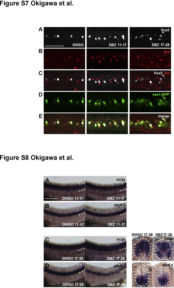
Fig. S6 DBZ-treatment affects V2 interneuron development. (A–E) V2a and V2b cells were identified as vsx1:GFP/Vsx2 double positive and vsx1:GFP/Scl double positive cells, respectively. Embryos were treated with DBZ either from 11 to 17 hpf (DBZ 11–17) or from 17 to 28 hpf (DBZ 17–28). Side views of embryos at 24 hpf with anterior to the left, dorsal up. Bar scale: 50 µm. Fig. S8: Reduction of p2 progenitor cells by17-28 hpf DBZ treatment. p2 progenitor cells detected by irx3a (C) and nkx6.1 (D) in embryos treated with DBZ at 17–28 hpf were slightly but significantly reduced compared to those treated at 11–17 hpf (irx3a (A) and nkx6.1 (B)) (70%, irx3a, n=10; 80%, nkx6.1, n=10). Left two panels: side views of embryos at 28 hpf with anterior to the left, dorsal up. Dorsal and ventral borders of the neural tubes are shown by the dotted lines. Bar scale: 50 µm. Right two panels (in C, D): transverse sections through the trunk region in embryos at 28 hpf. p2 Progenitor domains within the neural tubes (circled by white dotted lines) are indicated by red dashed outlines. Bar scale: 20 µm.
Reprinted from Developmental Biology, 391(2), Okigawa, S., Mizoguchi, T., Okano, M., Tanaka, H., Isoda, M., Jiang, Y.J., Suster, M., Higashijima, S.I., Kawakami, K., Itoh, M., Different combinations of Notch ligands and receptors regulate V2 interneuron progenitor proliferation and V2a/V2b cell fate determination, 196-206, Copyright (2014) with permission from Elsevier. Full text @ Dev. Biol.