
|
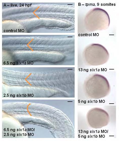
Fig. S2 Morpholino studies reveal that six1a and six1b function together during zebrafish myogenesis. Live images (A) at 24 hours post fertilization (hpf), anterior to the left, show that lower doses of six1a MO and six1b MO injected alone do not disrupt normal somite morphology individually, only when injected together are U-shaped somites observed (control: n=75/77, 6.5 ng six1a MO: n=61/64, 2.5 ng six1b MO: n=50/56, six1a/b MO: n=100/154). Lateral views of in situ hybridization (B) at 9 somites shows an early delay of tropomyosin-α (tpma) (control: n=25/27, six1a MO: n=21/25, six1b MO: n=20/25, six1a/b MO: n=10/14), which is strongest in the double six1a/b knockdown. Scalebar = 100 µm.