
|
Fig. 3
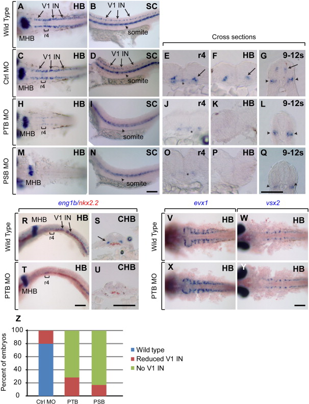
prdm12b is required for eng1b expression in the p1 domain. (A–Q) eng1b Expression in 24hpf wild type (A, B), control MO-injected (C–G), PTB MO-injected (H–L), PSB MO-injected (M–Q) embryos. (R–U) eng1b and nkx2.2 expression in 24 hpf wild type (R, S) and PTB MO-injected (T, U) embryos. (V, X) evx1 Expression in wild-type (V) and PTB MO-injected (X) embryos. (W, Y) vsx2 Expression in wild-type (W) and PTB MO-injected (Y) embryos. Embryos are shown in dorsal (A, C, H, M, V–Y) or lateral (B, D, I, N, R, T) view with anterior to the left, or in cross section (E–G, J–L, O–Q, S, U) with dorsal to the top. Sections were taken through r4, the hindbrain, the caudal hindbrain or the level of somites 9–12, as indicated. (Z) Quantification of eng1b phenotype. Scale bars are 100 μm. Brackets and asterisks indicate r4, arrows mark V1 interneurons and arrowheads mark somites. MHB – midbrain–hindbrain boundary, HB – hindbrain, CHB – caudal hindbrain, and SC – spinal cord.
Reprinted from Developmental Biology, 390, Zannino, D.A., Downes, G.B., Sagerström, C.G., prdm12b specifies the p1 progenitor domain and reveals a role for V1 interneurons in swim movements, 247-60, Copyright (2014) with permission from Elsevier. Full text @ Dev. Biol.