
|
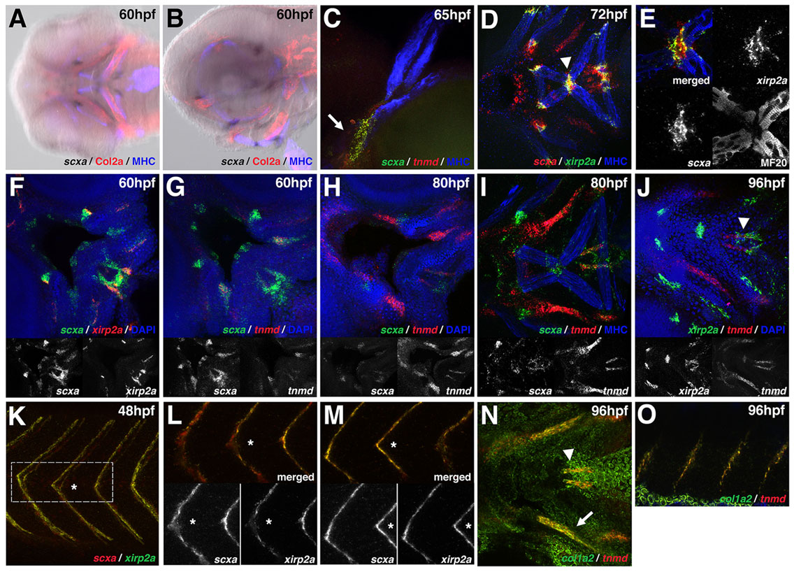
Fig. 2 Tendon genes mark discrete domains joining muscle and cartilage. (A,B) scxa is expressed at points of attachment of muscle-to-cartilage and cartilage-to-cartilage in the craniofacial tissue at 60hpf. Images were generated by overlaying the bright-field and fluorescent channels. (C) scxa and tnmd are co-expressed at the base of the cleithrum (arrow). (D-F) scxa and xirp2a are co-expressed in muscle attachment points, e.g. where the interhyoideus and intermandibularis muscles intersect (D, arrowhead; enlarged in E). Single channels of confocal image are shown in E. (G-I) Colocalization of scxa and tnmd is detected between 60 and 80hpf. Expression of scxa and tnmd transcripts is temporally dynamic: there is robust expression of scxa at 60hpf followed by weaker expression after 80hpf; tnmd is weakly expressed at 60hpf and increases in expression after 80hpf. (J) At 96hpf, xirp2a and tnmd are co-expressed in regions proximal to the muscle (arrowhead). (K-M) scxa and xirp2a are co-expressed in the tail myosepta in regions medial (L) and lateral (M) to the notochord. Single channels of confocal images are shown in L,M and an asterisk marks the corresponding myoseptum in the same embryo. (N,O) At 96hpf, tnmd and col1a2 are co-expressed in regions medial to the palatoquadrate (N, arrow), at the sternohyoideus connection point (N, arrowhead), and in the myosepta (O). Ventral (A,D-J,N) and lateral (B,C,K-M,O) views of flat-mounted embryos.