
Image
|
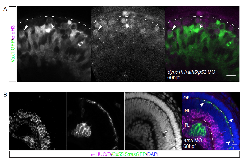
Figure Caption
Fig. S3
A) Apical division of Vsx1+ cells upon knockdown of ath5 and dync1h1, Related to Figure 4. Vsx1:GFP (green) transgenic embryos injected with a combination of morpholinos against dync1h1, ath5 and p53, stained for pH3 (magenta). Apical (arrow) and non-apical (notched arrow) divisions of Vsx1+ cells.
B) Horizontal cells inhabit the most basal layer in ath5 morphants, Related to Figure 5. Cx55.5:rasGFP (green) transgenic embryos injected with 4ng ath5 morpholino, stained with the AC/RGC marker α-HUC/D (magenta) and DAPI (blue). Arrows point towards Cx55.5+ HCs in the HC layer.
Acknowledgments
This image is the copyrighted work of the attributed author or publisher, and
ZFIN has permission only to display this image to its users.
Additional permissions should be obtained from the applicable author or publisher of the image.
Full text @ Cell Rep.