
|
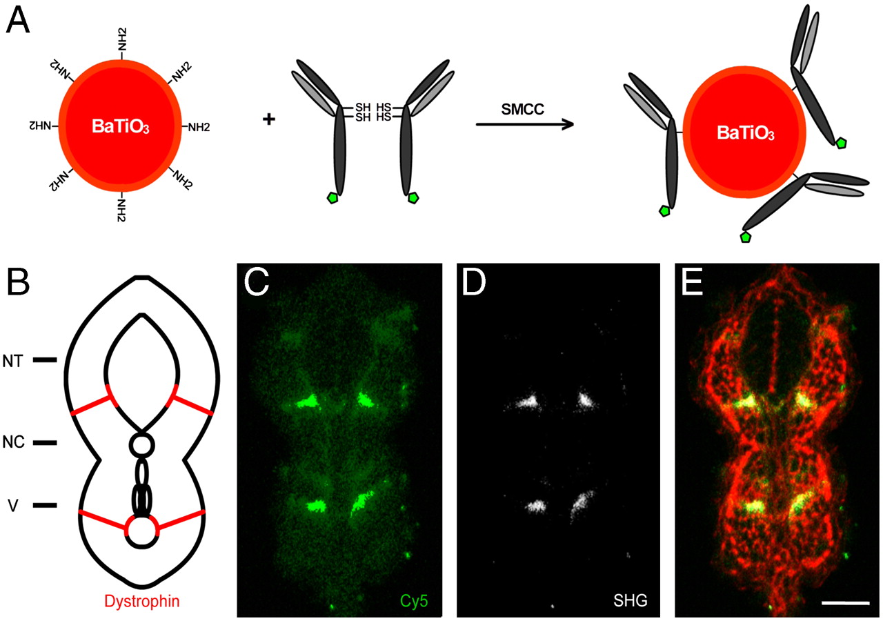
Fig. 5 SHG nanoprobe targeting specificity. (A) Scheme of SHG nanoprobe conjugation to Cy5-coupled (green pentagon) antibody fragments via disulphide reduction and sulfhydryl-amine coupling. (B) Schematic representation of a transversal section of trunk of a 24 hpf zebrafish embryo showing Dystrophin protein localization to fiber ends (red). NT, neural tube; NC, notochord; V, vessels. (C–E) Immunostaining showing Dystrophin protein localization in a transversal tissue section using secondary antibody coupled to Cy5 (green) and BaTiO3 (white). Both readouts—Cy5 immunofluorescence (C), and BaTiO3 SHG signal (D)—label specifically Dystrophin. (E) Phalloidin labeling (red) is superimposed to show cell profiles in the transversal tissue section. Note that SHG immunostaining provides superior SNR of Dystrophin detection using a narrow emission filter. Bar = 30 μm.