
|
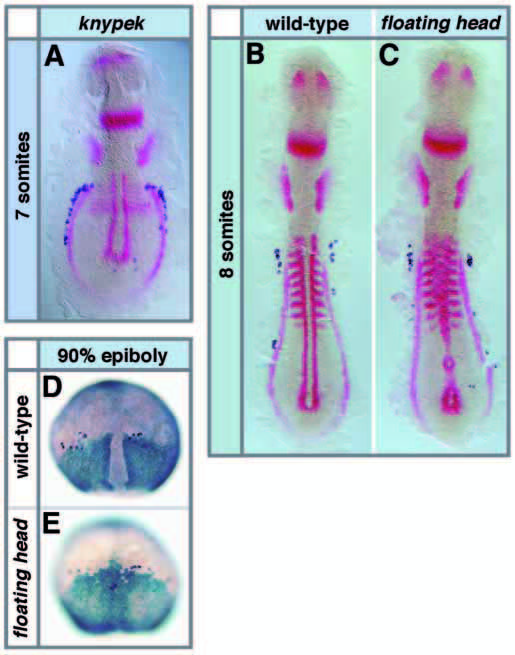
Fig. 6 PGC migration phenotype of knypek and floating head mutant embryos. All embryos were stained with vasa in dark blue and other probes in red or light blue as indicated. (A) Trailing PGCs align along the abnormally located pronephros (stained with pax2.1, adaxial cells and somites with myoD) in knypek mutant embryos at the 7- somite stage. (B,C) Normal arrangement of PGCs in wild-type (B) and floating head mutant (C) embryos at the 8-somite stage stained with myoD and pax2.1. (D,E) Dorsal view of embryos at the 90% epiboly stage showing that dorsally located PGCs do not migrate away from the dorsal midline in floating head mutant embryos. (D) In wild-type embryos, PGCs are not found at the region of the forming notochord marked by lack of papc staining, but they extend into the dorsal midline that expresses papc in floating head mutant embryos (E).