
|
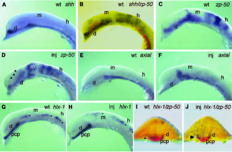
Fig. 4 Alterations of gene expression in 10-12 somite stage shh-injected embryos. Embryos injected (inj) with 25 ng/ml shh RNA (D,F,H,J) or control embryos (A,B,C,E,G,I) fixed at the 10-12 somite stage were analyzed by in situ hybridization. (A) shh staining in the ventral CNS. (B) The longitudinal expression domains of shh (red) and zp-50 (purple) occupy very similar territories in the ventral diencephalon. (C,D) zp-50 expression is dorsally expanded in the ventral diencephalon and the midbrain of shh-injected embryos. The beginning expression in the telencephalon (cf. B) is reduced in injected embryos. (E,F) axial expression is extended dorsally in the posterior diencephalon. (G,H) hlx-1 expression in the prechordal plate and the ventralmost midline ectoderm is not significantly altered by shh overexpression. However, expression (asterisks) in the mid- and hindbrain is shifted to more dorsal locations. (I,J) Crossections through the middiencephalon of double-labeled embryos show the dorsal expansion (arrowheads) of zp-50 expression (purple), whereas hlx-1 expression in the mesoderm and in the ventralmost ectodermal tissue is unaltered (red). d, diencephalon; h, hindbrain; m, mesencephalon; pcp, prechordal plate.