
Image
|
Figure Caption
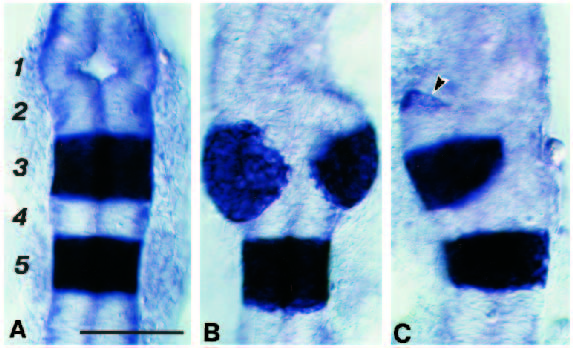
Fig. 6 Photomicrographs of the anterior hindbrain region of 20 h embryos hybridised with a probe for krx-20. (A) Control showing krx-20 expressing in r3 and r5. (B,C) Examples of embryos injected with mouse Hoxa-1 RNA at the single cell stage. r5 is normal in both cases but r3 expression of krx-20 is abnormal. The anterior hindbrain appears distorted; thus r3 is split dorsally in B and is laterally displaced in C. Ectopic regions of krx-20 expression, such as that arrowed most anteriorly in C, were occasionally seen. Scale bar, 100 μm.
Figure Data
Acknowledgments
This image is the copyrighted work of the attributed author or publisher, and
ZFIN has permission only to display this image to its users.
Additional permissions should be obtained from the applicable author or publisher of the image.
Full text @ Development