
|
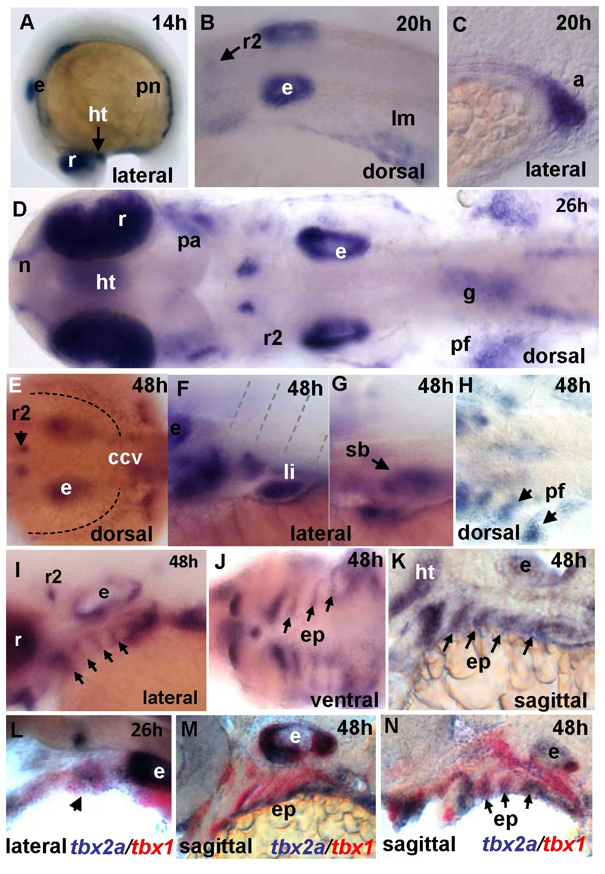
Fig. 1
Expression pattern of tbx2a during development as detected by whole-mount in situ hybridization (WISH) (14-48 hpf).
(A) Lateral view of 14 hpf embryo. Lateral-dorsal view of 20 hpf embryo (B) hindbrain and (C) lateral view of the anus. (D) Composite image showing a dorsal view of 26 hpf embryo. (E-K) 48 hpf embryo. (E) Dorsal view of the hindbrain. (F) Lateral view at the level of somite 2. (G) Lateral view of the swim bladder. (H) Dorsal view of the pectoral fins. (I-K) Pharyngeal arches in a (I) ventral view and in (J-K) sagittal sections. Two-color WISH for (K) dlx2 (magenta) and tbx2a (red), (L-N) tbx2a (magenta) and tbx1 (red). Abbreviations for all figures: a: anus; pa: pharyngeal arches; ccv: common cardinal vein; e: ear; ep: endodermal pouch; g: gut; r: retina; r2: rhombomere 2; ht: hypothalamus; h: hours post-fertilization; li: liver; lm: lateral mesoderm; n: nasal pits; ncc: neural crest cells; pf: pectoral fin; pn: pronephric ducts; sb: swim bladder; v: vagal nucleus.