
Image
|
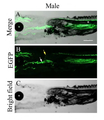
Figure Caption
Fig. S3 Origin of BT vascularization. Longitudinal sections of the area connecting the body to the pectoral fin of males (A-C) of the Tg(fli1a:EGFP) fish. A blood vessel (yellow arrow) is observed sprouting from the central artery (white arrow) and projecting parallel to the hemiray (white asterisk). (A) Merge = Brightfield + GFP; (B) GFP fluorescence; (C) Brightfield. Scale bars = 100μm (shown in A).
Acknowledgments
This image is the copyrighted work of the attributed author or publisher, and
ZFIN has permission only to display this image to its users.
Additional permissions should be obtained from the applicable author or publisher of the image.
Full text @ Development