
|
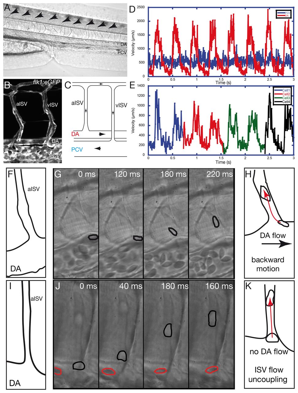
Fig. 1 Blood cell motions demonstrate flow rectification and an additional force at the ISV entry of the embryonic vascular network. (A) Lateral view of the zebrafish larvae at 72 hpf. Blood vessels of the tail are visible in the focal plane. Blood cells are present in the dorsal aorta (DA) and the posterior cardinal vein (PCV). Arrows indicate the intersegmental vessels (ISV). (B) View of a typical vascular loop between the DA and the PCV through z projection of a confocal stack obtained with the Tg(flk1:eGFP) transgenic line expressing the EGFP specifically in the endothelial cells. Arterial ISV (aISV) and venous ISV (vISV) are labeled. (C) A typical vascular loop observed in the trunk vasculature with the black arrows indicating the direction of blood flow. The blood cells in the DA enter into the aISV, pass in to vISV and move towards the PCV and the heart. (D) Typical velocity profile of the blood cells circulating in the DA (red) and PCV (blue). The DA displays a pulsatile flow, whereas the PCV flow is relatively steady. (E) Typical velocity pattern of blood cells observed in the ISV. Flow is pulsatile but not fully synchronized with the DA flow. (F-K) Fast imaging of blood cell motion at the ISV entry. (F) The observed topography at the ISV entry in G. (G) Fast time-lapse imaging showing a blood cell (outlined in black) moving against the DA flow towards the ISV. (H) Summary of the backward blood cell motion towards the ISV (red arrow) as opposed to DA flow (black arrow). (I) The observed topography at the ISV entry in J. (J) Fast time-lapse imaging showing blood cell displacement in the ISV (outlined in black) is much greater than in the DA (outlined in red). (K) Summary of the additional cell motion within the ISV (red arrow) when there is no visible DA flow.