
|
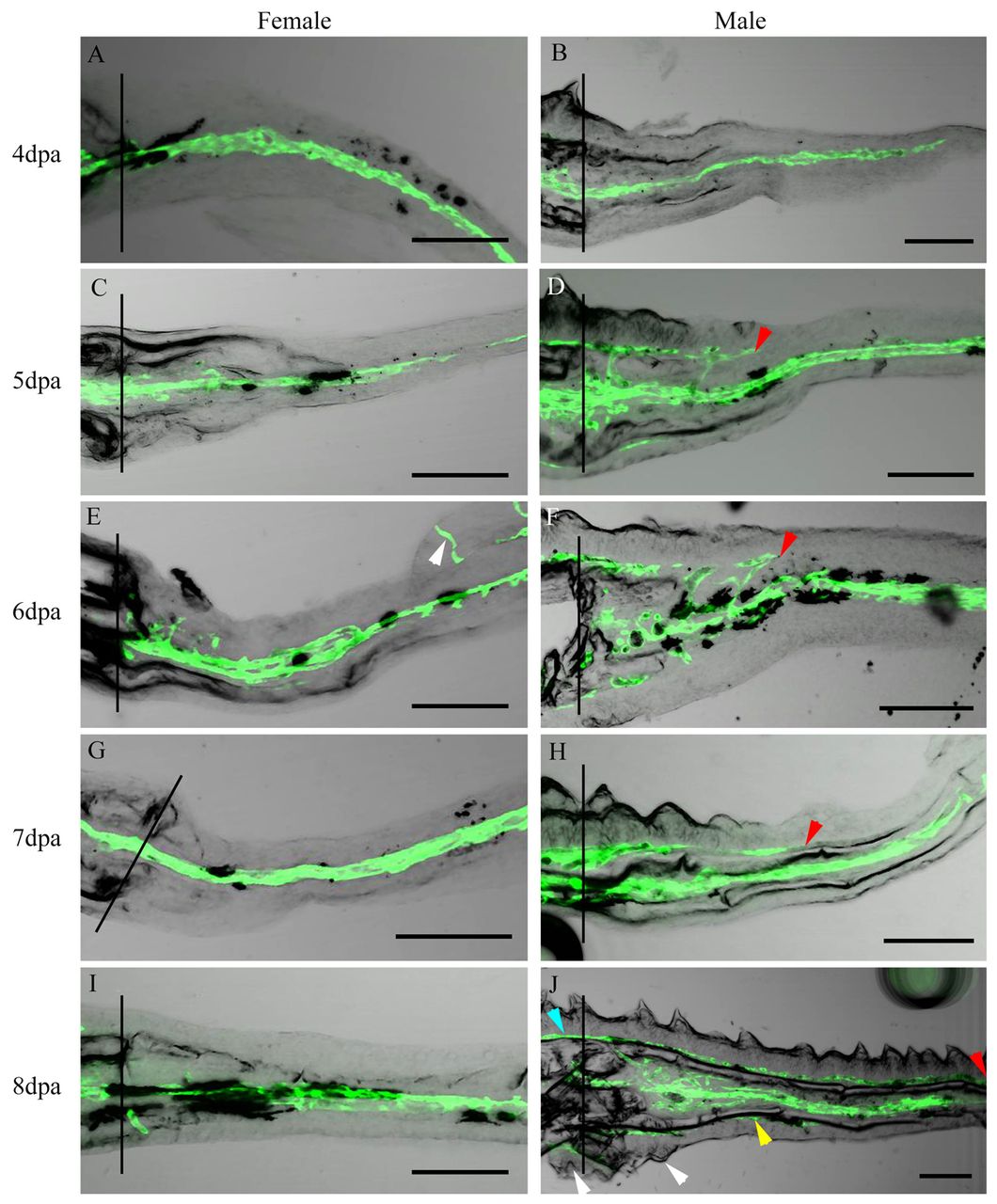
Fig. 5 BT cluster regeneration occurs alongside a second wave of neo-angiogenesis. Confocal images of longitudinal sections of regenerating male and female pectoral fins. (A,C,E,G,I) Female blood vessels regenerate along the center of the ray. (E) Arrowhead indicates the distal ray folded back on itself. (B) The first wave of male blood vessel regeneration occurs down the center of the ray. (D,F,H,J) At 5-8 dpa, a second wave travels in a proximal-to-distal fashion along the base of the epidermis (red arrowhead). (J) New BTs (white arrowheads) and blood vessels (yellow arrowhead) are observed in the ventral fin regenerate. Blue arrowhead indicates blood vessels along the base of BTs located in the stump. Black vertical lines indicate the amputation plane. Scale bars: 100 μm.