
Image
|
Figure Caption
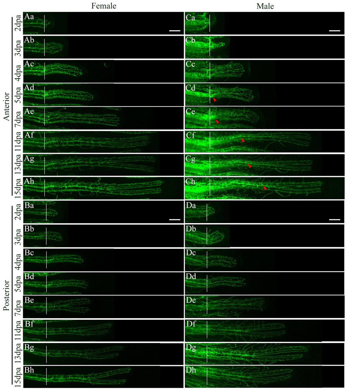
Fig. 4 Male and female Tg(fli1a:EGFP) blood vessel regeneration. (Aa-h,Ba-h,Da-h) Blood vessel regeneration follows similar patterns in the female (A,B) and posterior (D) male rays. (Ca-h) Plexus formation (Cb) is delayed in the male anterior ray compared with females (Ab) and appears irregular in shape. (Cd-h) At 5 dpa, a second wave of neo-angiogenesis occurs underneath new BT formation in the male anterior ray and proceeds in a proximal-to-distal manner (red arrowheads). Anterior, fin ray 4; posterior, fin ray 7. White lines indicate the plane of amputation. Scale bars: 100 μm.
Acknowledgments
This image is the copyrighted work of the attributed author or publisher, and
ZFIN has permission only to display this image to its users.
Additional permissions should be obtained from the applicable author or publisher of the image.
Full text @ Development