
Image
|
Figure Caption
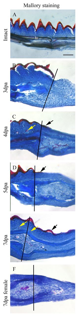
Fig. 3 Male pectoral fin BT regeneration. Mallory stain of intact (A) and regenerating (B-F) pectoral fin longitudinal sections. (A,B) Keratinized caps (red) and underlying epidermis (blue) are observed on intact fins (A) but are lost in 3-dpa regenerates (B). (C,D) At 4-5 dpa, a layer of keratin is deposited in the proximal regenerate (black arrows). (E) At 7 dpa, BTs (black arrow) are observed in the fin regenerate. (F) Females do not possess BTs. Yellow arrows indicate blood cells. Black lines (B-F) indicate the amputation plane. dpa, days post-amputation. hr, hemiray. Scale bar: 50 μm.
Acknowledgments
This image is the copyrighted work of the attributed author or publisher, and
ZFIN has permission only to display this image to its users.
Additional permissions should be obtained from the applicable author or publisher of the image.
Full text @ Development