
|
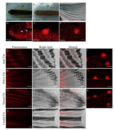
Fig. S1 Distribution and morphology of BTs in male zebrafish. (A) Dorsal view of a male zebrafish with pectoral fin BTs (black arrowhead). (B) Dorsal view of a female zebrafish with no BTs and translucent fins (black arrowhead). (C) Pectoral fin of a male longfin mutant with long BT clusters (outlined in blue). (D) Grouped BTs in a line/semi circle (white box) and isolated BTs (white arrowheads) on the side of a male zebrafish head. (E) BT on a barbel. (F) A BT on a scale pulled from the body surface. (G-J) BTs distributed along the anal fin rays. (K-N) BTs along the pelvic fin rays. (O-R) BTs along the dorsal fin rays. (S-U) No BTs are observed on the caudal fin. Fluorescence Tg(KR21) images (red), Merged (Tg(KR21) + Brightfield) images. Scale bar for C = 200μm; D = 400μm; E = 12.5µm; J, N, R, F = 25μm; G-I, K-M, O-Q, S-U = 200μm.