
|
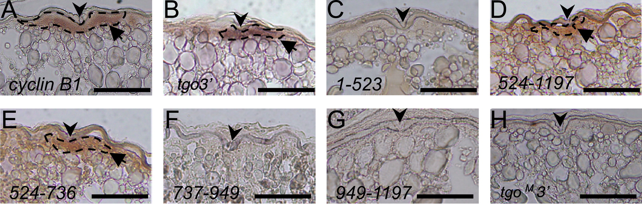
Fig. 4 Localization of cyclin B1 reporter mRNAs in the animal polar cytoplasm of transgenic zebrafish oocytes. (A) Section in situ hybridization of a full-grown oocyte probed with cyclin B1. The cyclin B1 mRNAs were localized to the animal polar cytoplasm of the oocyte beneath the micro-pile as an aggregation (arrow). ((B)–(H)) Section in situ hybridization probed with gfp, showing full-grown oocytes of transgenic zebrafish expressing tgo3′ (B), 1–523 (C), 524–1197 (D), 524–736 (E), 737–949 (F), 949–1197 (G) and tgoM3′ (H) mRNAs. The tgo3′, 524–1197 and 524–736 mRNAs were aggregated beneath the micro-pile (arrows) ((B), (D), (E)), whereas the 1–523, 737–949, 949–1197 and tgoM3′ mRNAs had no signal ((C), (F), (G), (H)). Arrowheads indicate the micro-pile. Dotted lines encircle aggregated mRNAs. Bars, 100 μm.
Reprinted from Developmental Biology, 382(2), Yasuda, K., Kotani, T., and Yamashita, M., A cis-acting element in the coding region of cyclin B1 mRNA couples subcellular localization to translational timing, 517-29, Copyright (2013) with permission from Elsevier. Full text @ Dev. Biol.