
IMAGE
Fig. S5
- ID
- ZDB-IMAGE-131218-13
- Publication
- Sittaramane et al., 2013 - The PCP protein Vangl2 regulates migration of hindbrain motor neurons by acting in floor plate cells, and independently of cilia function
- All Figures
- Figures for Sittaramane et al., 2013
Image
|
Figure Caption
Fig. S5
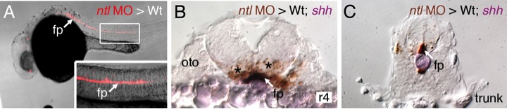
Targeting of transplanted donor cells to the floor plate of host embryos.
(A) Lateral view of a live 24 hpf wildtype embryo with donor-derived no tail (ntl) morphant cells (red, rhodamine and biotin dextran) in the floor plate. The embryo was then processed for shh in situ (purple) to label the floor plate and streptavidin immunochemistry (brown) to detect donorderived cells. Cross sections at the r4 (B) and trunk (C) levels show the presence of donor cells (arrows) in the floor plate. oto, otic vesicle. Asterisks in B indicate labeled cell debris from the transplantation procedure.
Acknowledgments
This image is the copyrighted work of the attributed author or publisher, and
ZFIN has permission only to display this image to its users.
Additional permissions should be obtained from the applicable author or publisher of the image.
Reprinted from Developmental Biology, 382(2), Sittaramane, V., Pan, X., Glasco, D.M., Huang, P., Gurung, S., Bock, A., Li, S., Wang, H., Kawakami, K., Matise, M.P., and Chandrasekhar, A., The PCP protein Vangl2 regulates migration of hindbrain motor neurons by acting in floor plate cells, and independently of cilia function, 400-412, Copyright (2013) with permission from Elsevier. Full text @ Dev. Biol.