
Fig. 6
|
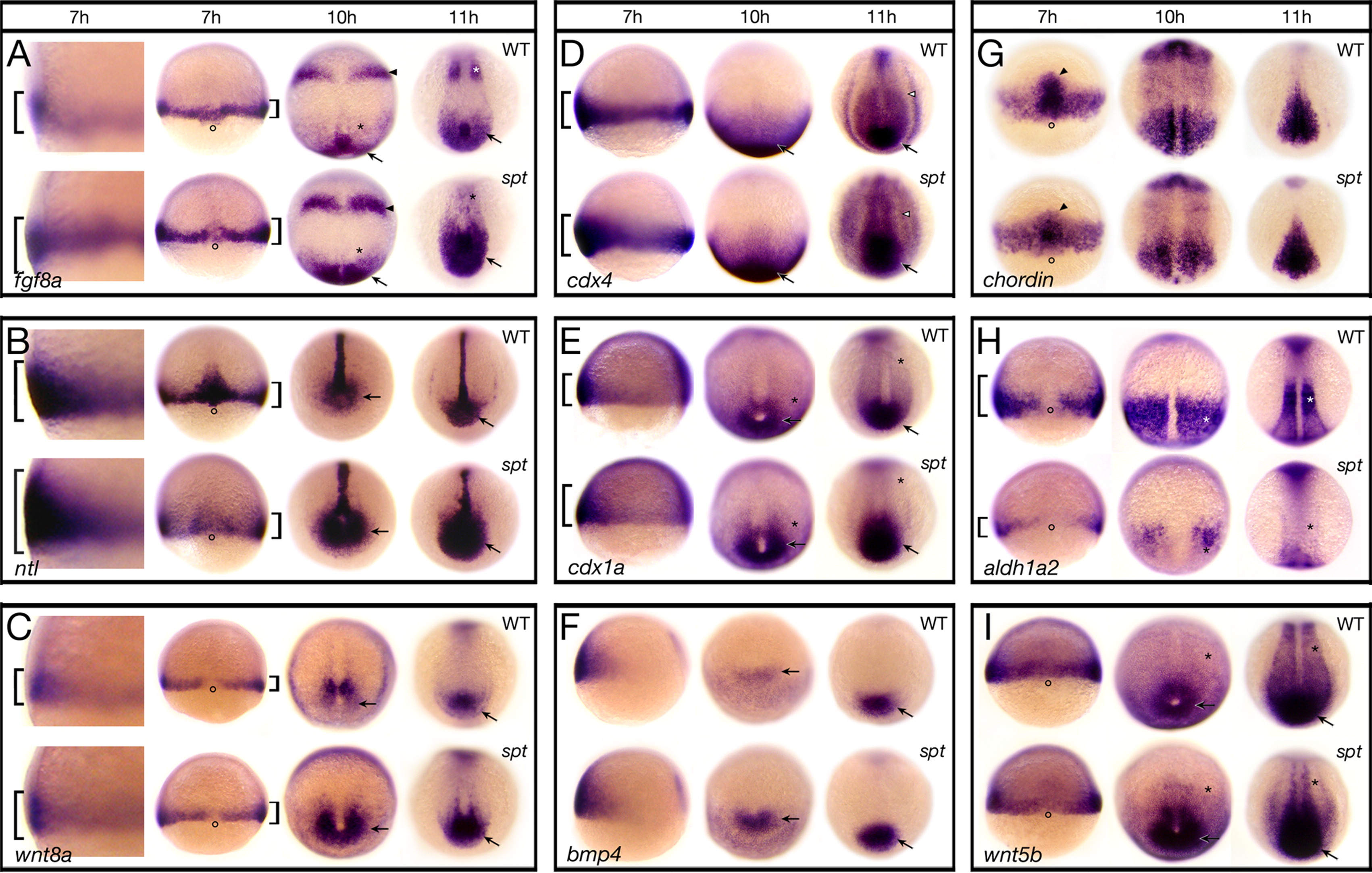
Fig. 6 Components of the Fgf and Wnt pathway are more highly expressed in spt mutants. Expression of (A) fgf8a; (B) ntl; (C) wnt8a; (D) cdx4; (E) cdx1a; (F) bmp4; (G) chordin; (H) aldh1a2; and (I) wnt5b at: 7 h, 10 h and 11 h. Embryos at 7 h are shown in (A–C) a high magnification left side view and a low magnification dorsal view, (D–F) a left side view, and (G–I) a dorsal view. Older embryos are all shown in a dorsal posterior view. Designations (7 h): brackets, more or less expression; open circles, dorsal side; arrowhead, prechordal plate; (later stages): arrow, more expression in the posterior region and tailbud; asterisk, less expression in the paraxial mesoderm; arrowhead, more expression in the future midbrain-hindbrain boundary; open arrowhead, more expression in the intermediate mesoderm.
Reprinted from Developmental Biology, 383(1), Warga, R.M., Mueller, R.L., Ho, R.K., and Kane, D.A., Zebrafish Tbx16 regulates intermediate mesoderm cell fate by attenuating Fgf activity, 75-89, Copyright (2013) with permission from Elsevier. Full text @ Dev. Biol.