
|
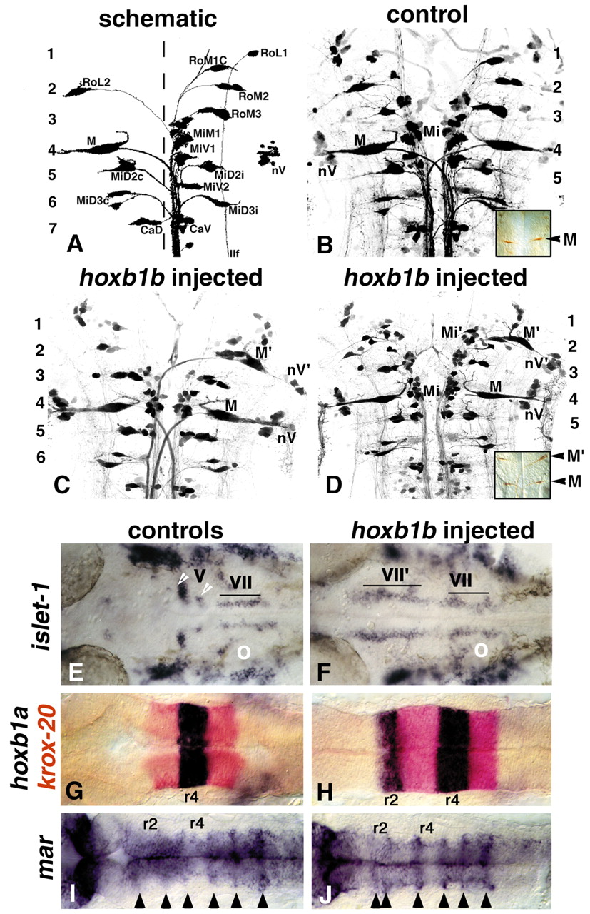
Fig. 5
Disposition of neuroanatomical and molecular markers reveals that mis-expression of hoxb1b transforms r2 to an r4 phenotype. (A-D) Retrograde labelling from the spinal cord of 5-day-old larvae reveals the disposition of the RS neurones (anterior to the top). (A) The subset of neurones labelled from the right hand side of the spinal cord only is schematized and labelled: M, Mauthner; nV, nucleus of the vestibular formation; MiM1, middle medial 1 cells; MiV1, middle ventral 1 cells; RoL2, rostral lateral 2 cells; llf, lateral longitudinal fascicle. Other abbreviations as previously designated (Hanneman et al., 1988). The locations of the rhombomeres relative to the RS neurones are indicated on the left-hand side. (B) Retrograde labelling of a normal 5-day-old larva. Mi, MiM1 plus MiV1 cells; rhombomeres numbered on right-hand side. Inset, 3A10 antibody staining reveals the r4 Mauthner neurones, M. (C,D) Mis-expression of hoxb1b leads to formation of the r4 characteristic M, Mi and nV cells at the r2 level; ectopic neurones are indicated by 2. These examples show unilateral duplications, less frequently bilateral duplications were observed, as shown in the inset in (D) by 3A10 antibody staining to reveal ectopic Mauthner neurones at the r2 level, M2. (E-J) Whole-mount in situ hybridization analysis of injected embryos, mounted with dorsal side uppermost and anterior to the left; in each case an unmanipulated control embryo is shown on the left-hand side and a hoxb1b-injected embryo at the same stage on the right-hand side. (E,F) 28 h; (G-J) 19 h; (E) islet1 expression labels cell bodies of the branchiomotor neurones. The trigeminal (Vth nerve) cell bodies have a lateral location, show intense labelling, and are subdivided into a major anterior (r2) population and a minor posterior (r3) population (white arrowheads). The facial (VIIth nerve) cell bodies lie more medially, show lower level expression, and form an anterior-posterior array through r4-r6 at this stage (bracket). (F) hoxb1b-injected embryo, islet1 expression shows medial facial-like neurones (VII2) at the level of r2 and r1, and possibly extending into the midbrain. O, otic vesicle. (G) In wild-type embryos, hoxb1a (blue) is expressed in r4 and krox-20 (red) in r3 and r5. (H) In hoxb1b-injected embryos there is ectopic hoxb1a (blue) expression at the r2 level, note expansion of the r3 krox-20 territory (red). (I) mar is expressed at elevated levels in rhombomere boundaries (arrowheads). (J) In hoxb1b-injected embryos, mar boundary staining reveals expansion of the r3 territory at the expense of the r2 territory.