
|
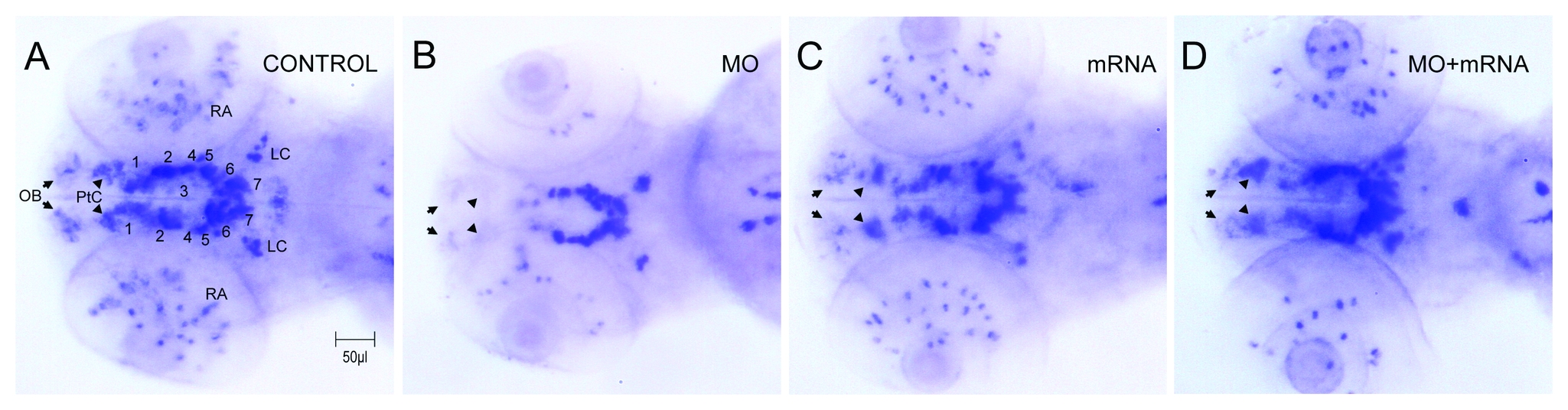
Fig. 6
Morphological changes in DA neuron patterning.
In situ hybridization with tyrosine hydroxylase anti-sense mRNA probe. (A) Control. (B) 5 ng ddc tMO1 treated. (C) 100 pg ddc mRNA treated. (D) Co-injected mixture with 5 ng ddc tMO1 and 50 pg ddc mRNA with non-tMO1 binding site. Numbers show the site of dopaminergic neuron clusters 1-7 and 8, which indicated spots in retina, locus coeruleus (LC), pre-tectum (PrC, arrowhead) and olfactory bulb (OB, arrow). Compared to tyrosine hydroxylase expression pattern in control larvae, several tissue parties including olfactory bulb, pre-tectum and DA cluster were malpositioned or absent in the ddc morphant brains.