
|
Fig. 6
Metastasis assay of zebrafish casper embryos transplanted with PaTu-S and PaTu-T cells.
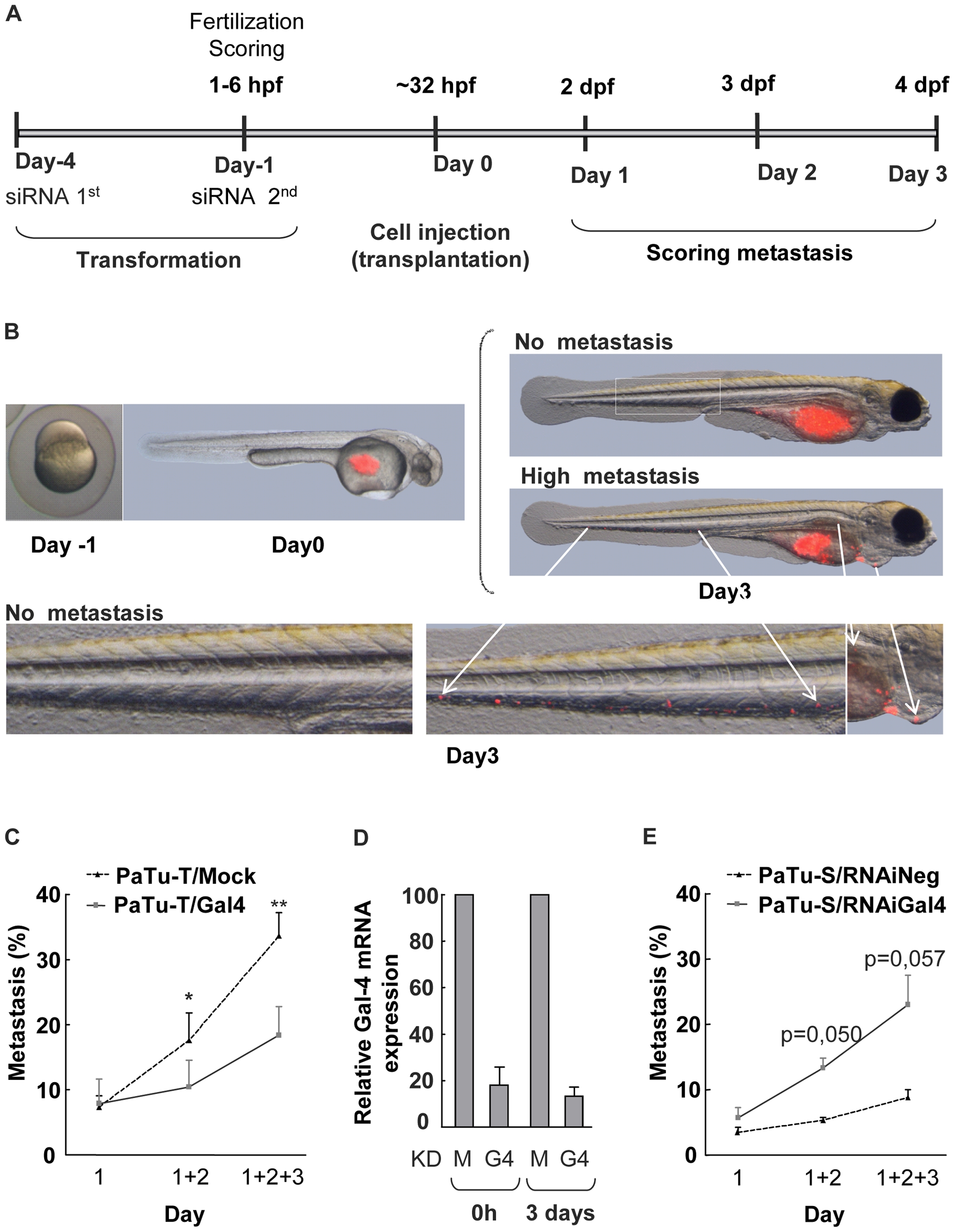
A) Schematic depiction of the time schedule of the transplantation experiments. Embryos were injected at the yolk sack at 32–38 h developmental stage. siRNA was introduced twice at day -4 and day -1, respectively. At 1–6 hours post fecundation (hpf) the embryos were evaluated for viability and fitness. Scoring of metastasis formation was performed at day 1, 2 and 3 by detection of the localization of CMDiI (red) fluorescent cells. B) Representative photographs of a casper embryo at one cell developmental stage, and an embryo injected with fluorescent red cells. At day 0 cells are present only at the yolk sac of the embryo and at day 3 cells had migrated from the yolk sac throughout the embryo, including the caudal vein, hart and liver. C) Metastasis assay of zebrafish casper embryos transplanted with PaTu-T/Gal-4 and PaTu-T/mock cells. The number of embryos presenting metastasis is shown per day and the total percentage of embryos with metastasis formation after 3 days is depicted as a histogram. The data are derived from 4 separate experiments, and are depicted as the average metastasis formation ± SEM. D) Gal-4 mRNA levels of PaTu-S/Gal-4 KD (KD G4) and mock siRNA treated (KD M) PaTu-S cells were determined by quantitative RT-PCR as a control for the efficiency of the siRNA treatment. E) Histogram showing the total percentage of embryos, transplanted with fluorescent PaTu-S/mock-KD and PaTu-S/Gal-4-KD cells, with metastasis formation after 1–3 days. Data are derived from 3 separate experiments, and are depicted as average metastasis formation ± SEM. Significance of the data is determined by paired sample T-tests (* pd0.05, ** pd0.01 and, *** pd0.001).