
|
Fig. 5
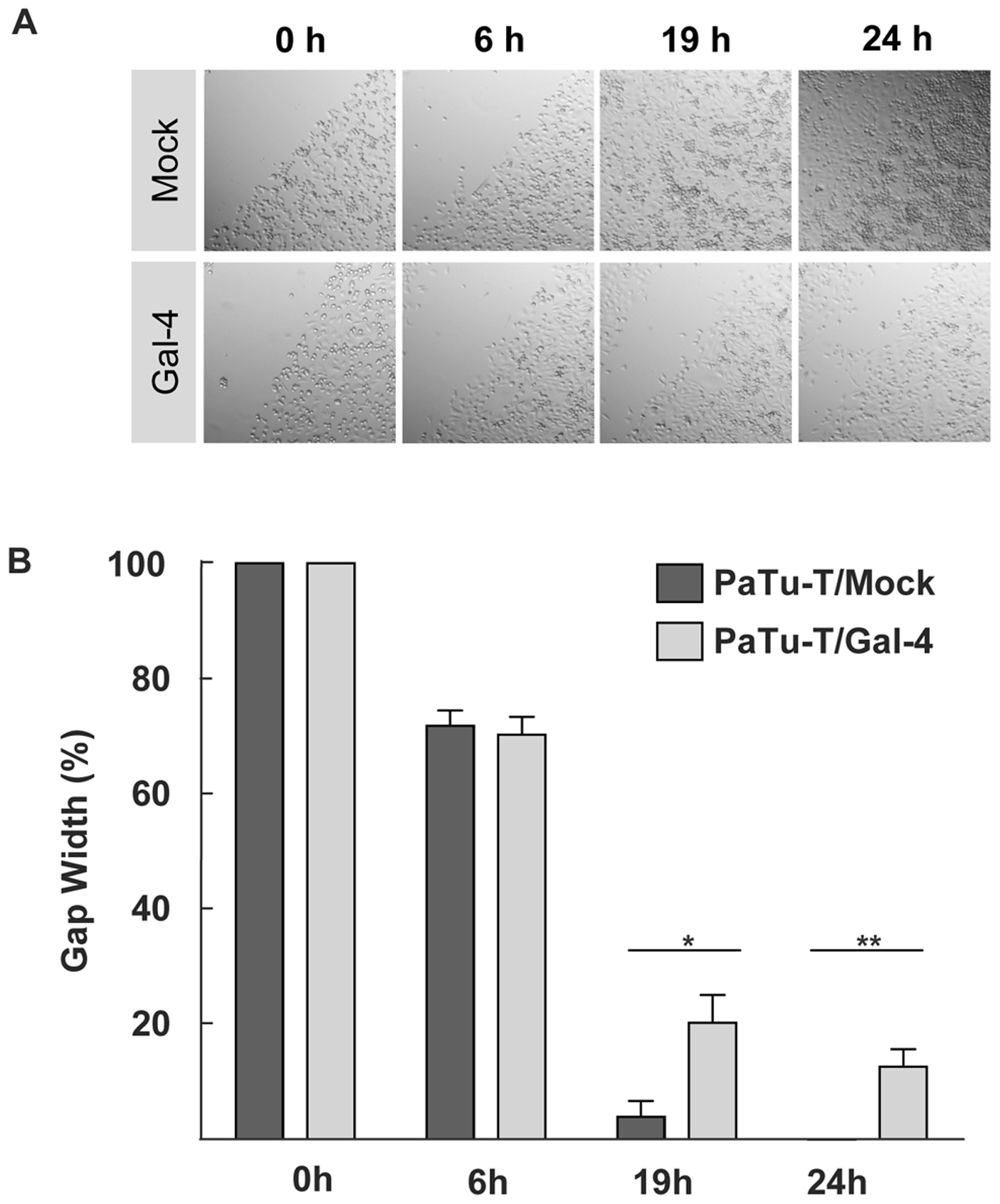
In vitro cell migration of PaTu-T cells.
A scratch (wound healing) assay was performed with PaTu-T, PaTu-T/Gal-4 and PaTu-T/mock cells. PaTu-T/mock and PaTu-T/Gal-4 cells were seeded on a 24 well plate and scratched on the surface with a 200-μl pipette tip. Relative values were set at 100% of the gap width at the time of the scratch. A) Representative photographs at time points 0, 6, 19 and 24 hours after the wound (scratch) for all conditions are depicted. B) Histogram representation of data analyzed from photographs taken at 0 h; 6 h, 19 h and 24 h after the scratch. Measurements were done in duplicate in 3 separate experiments, and data are depicted as average gap width ± SEM. (* pd0.05 and ** pd0.01, using one way ANOVA Tukey t tests).