
Image
|
Figure Caption
Fig. S3
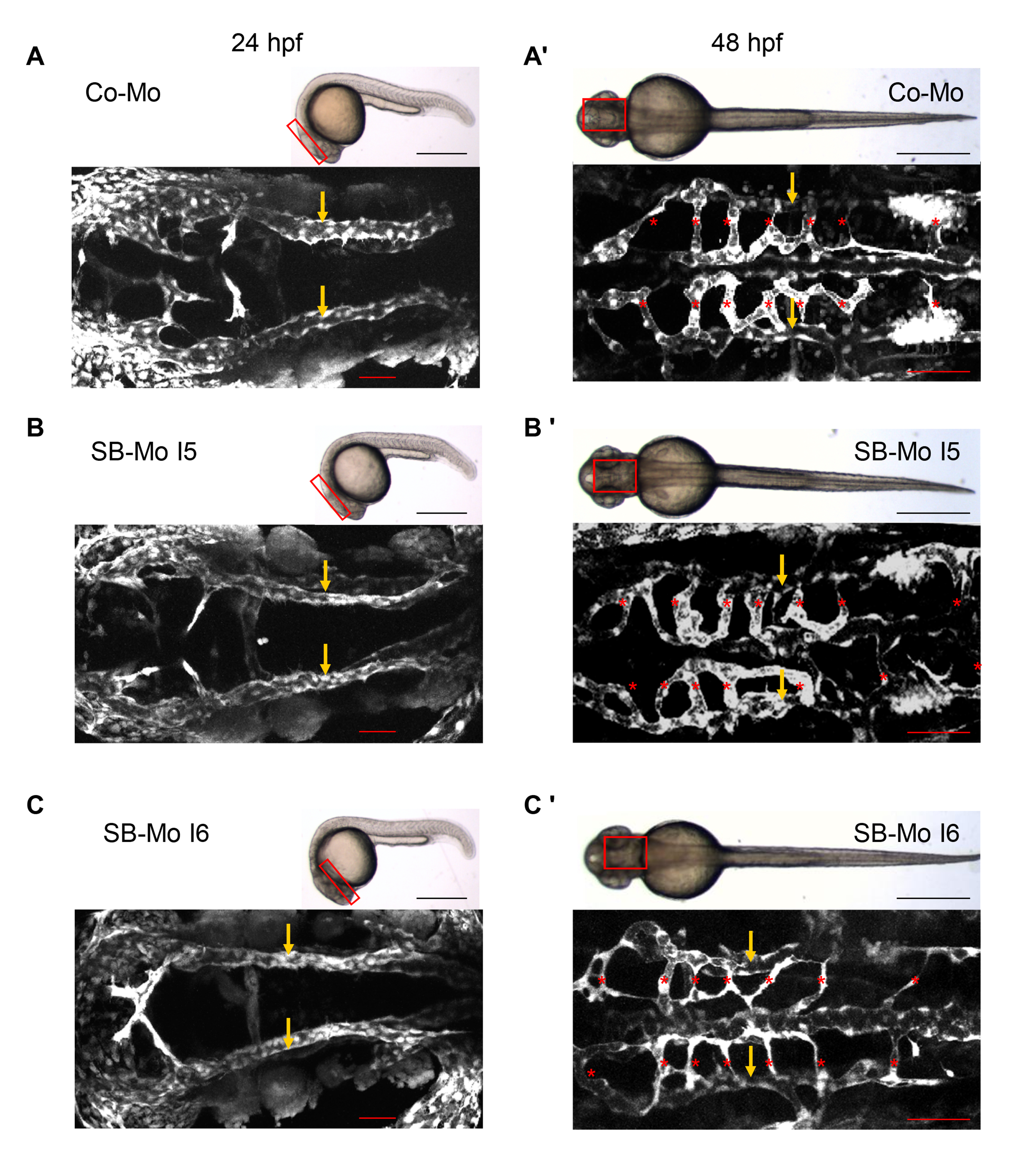
PKD1 silencing in zebrafish did not alter cranial angiogenesis. A–C2, 24 hpf (A, B, C) and 48 hpf (A2, B2, C2) tg(fli1:EGFP) zebrafish embryos were analyzed for defects in the primordial hindbrain channel (PHBC, arrows) and central arteries (CA, asterisks) by confocal microscopy. Injection of 500 pg SB-Mo I5 (B, B2) or 500 pg SB-Mo I6 (C, C2) did not reveal vascular defects in the cranial vasculature as compared to 2 ng Co-Mo injected tg(fli1:EGFP) zebrafish embryos (A, A2). Black scale bars: 500 μm, red scale bars: 50 μm.
Acknowledgments
This image is the copyrighted work of the attributed author or publisher, and
ZFIN has permission only to display this image to its users.
Additional permissions should be obtained from the applicable author or publisher of the image.
Full text @ PLoS One