
|
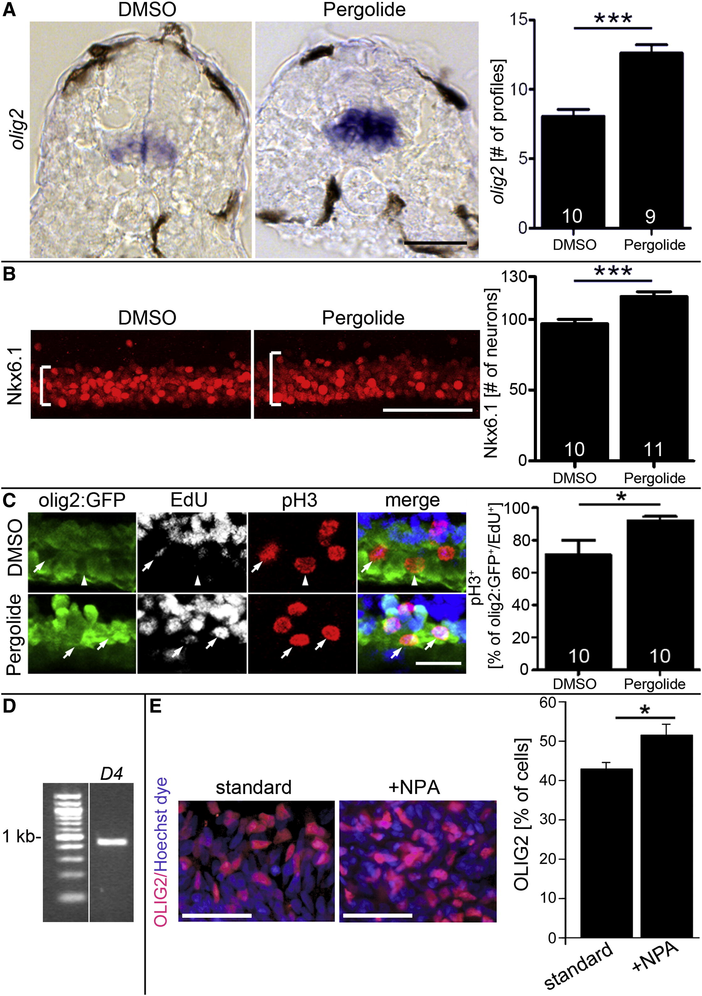
Fig. 4 Dopamine Agonists Affect Ventral Progenitor Cells (A) The olig2 mRNA expressing pMN progenitors are increased in number and labeling intensity after pergolide treatment (24–33 hpf; cross-sections; p < 0.0001). (B) Nkx6.1 labeling is expanded (brackets) after pergolide treatment (24–33 hpf; lateral views; p < 0.0001). (C) A higher proportion of pMN progenitors in M phase (olig2:GFP+/pH3+) is labeled with EdU 2 hr after exposure, indicating shortened G2 phase, after pergolide treatment (24–33 hpf; lateral views; p < 0.05; arrows indicate triple-labeled cells, and arrowhead indicates a cell that is olig2:GFP+/pH3+/EdU-). (D and E) In human embryonic stem cell cultures, driven toward motor neuron differentiation, PCR indicates expression of the d4 receptor at day 16 (D) and the proportion of OLIG2+ cells is significantly (p = 0.0109) increased by NPA at day 16 (E). Error bars represent SEM. Scale bars, 50 μm (A, B, and E) and 20 μm (C). See also Figure S4.
Reprinted from Developmental Cell, 25(5), Reimer, M.M., Norris, A., Ohnmacht, J., Patani, R., Zhong, Z., Dias, T.B., Kuscha, V., Scott, A.L., Chen, Y.C., Rozov, S., Frazer, S.L., Wyatt, C., Higashijima, S., Patton, E.E., Panula, P., Chandran, S., Becker, T., and Becker, C.G., Dopamine from the Brain Promotes Spinal Motor Neuron Generation during Development and Adult Regeneration, 478-491, Copyright (2013) with permission from Elsevier. Full text @ Dev. Cell