
|
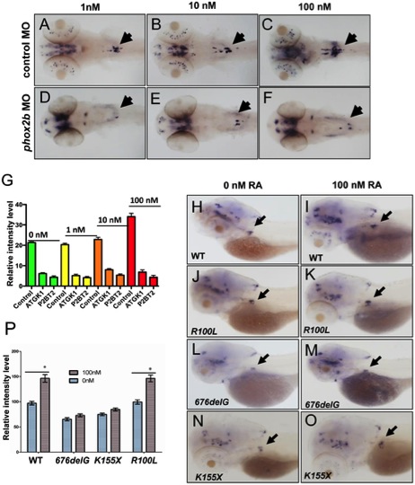
Fig. 4 Impaired differentiation in the SCG due to phox2b deficiency is not rescued by retinoic acid.
(A–F) Dorsal views of 3-dpf embryos injected with phox2b MO (D–F) or mismatched control MO (A–C) and treated with increasing concentrations of 13–cis retinoic acid (RA). (G) Relative intensity measurements of th expression in the SCG of embryos injected with various MOs and treated with different concentrations of RA. ATGK1, translation-blocking phox2b MO; P2BT2, splice blocking phox2b MO. (H–O) Whole-mount ISH of 3-dpf embryos in which the specified RNAs were overexpressed and analyzed for th expression following exposure to RA. Capped mRNA (100 ng/µl) for wild-type (WT) human PHOX2B, and the R100L, 676delG, K155X mutations were injected into embryos at the one-cell stage. (P) Quantification of the relative intensity of th staining in the embryos depicted in H–O. Data are presented as means ± SD (*P<0.05; n = 10 per group).